What Are Peptides? A Beginner's Guide
Peptides are short chains of amino acids found naturally in your body that regulate everything from metabolism to immunity. This beginner's guide explains what peptides are, how they work, the major categories used in health research, how to evaluate the evidence, and how to navigate the regulatory landscape before exploring peptide-based therapies.
Medical Disclaimer: This article is for informational and educational purposes only. Peptides discussed here are not FDA-approved for all uses. Nothing in this article constitutes medical advice. Always consult a qualified healthcare provider before considering any peptide therapy or investigational compound.
If you’ve been reading about biohacking, longevity clinics, weight loss medications, or sports medicine, you’ve encountered the word “peptide” — probably attached to bold claims about fat loss, healing, anti-aging, or performance. Some of those claims are backed by solid clinical trial data. Many are extrapolations from animal research. Some are marketing.
Knowing which is which requires understanding what peptides actually are, how they work biologically, why synthetic versions have become a significant category of both pharmaceuticals and research compounds, and what framework to use when evaluating any specific peptide claim.
This guide covers all of that from the ground up. No biochemistry degree required.
What Is a Peptide?
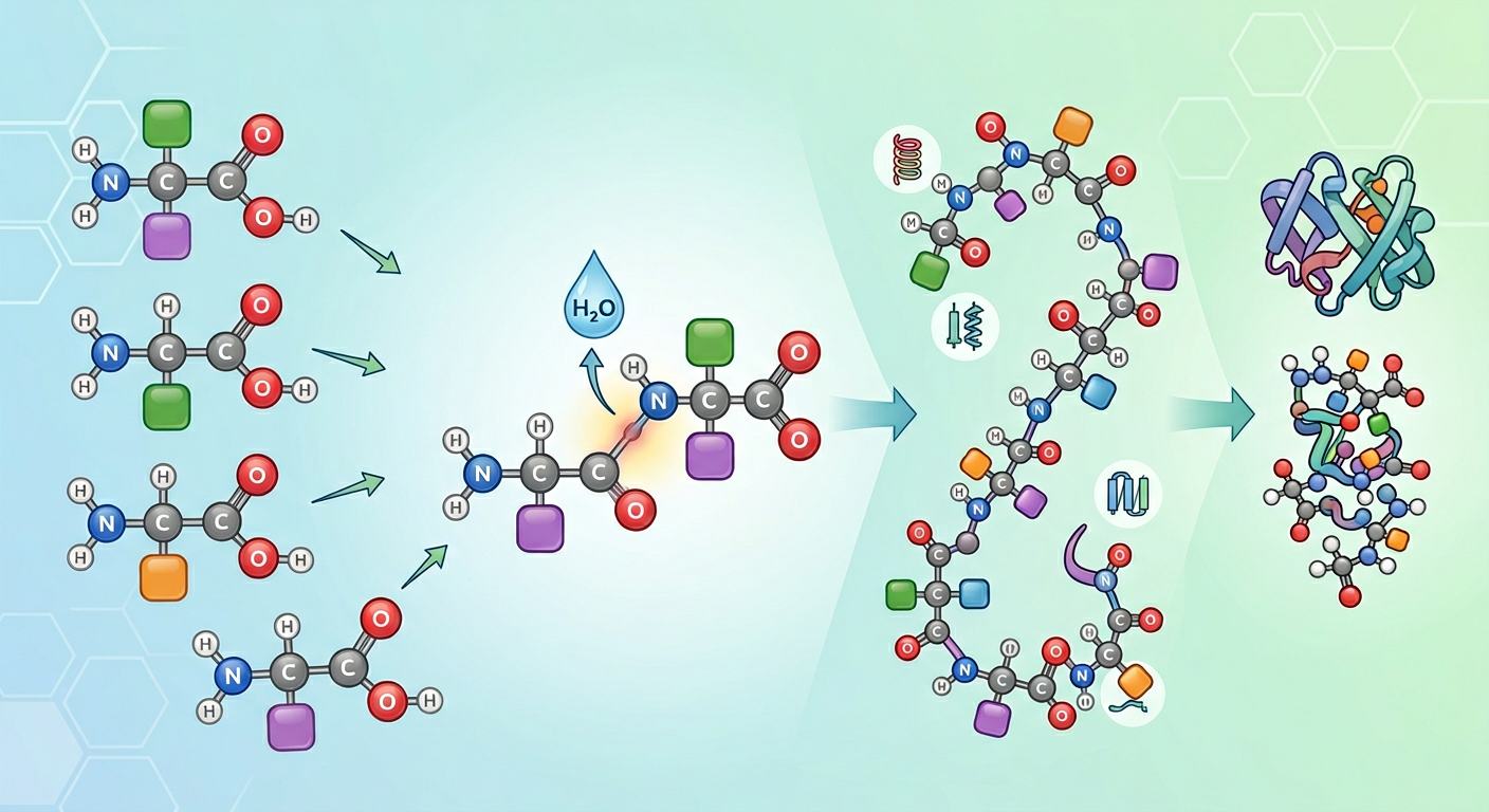
A peptide is a short chain of amino acids linked together by peptide bonds — the chemical connections between the amino group of one amino acid and the carboxyl group of the next. Amino acids are the molecular building blocks of all proteins, and when they join in chains, they form either peptides (shorter chains) or proteins (longer chains).
The boundary between a peptide and a protein is not hard: the convention is roughly:
- Amino acids: individual building blocks (20 standard ones, plus several less common)
- Peptides: chains of 2–50 amino acids
- Proteins: chains of 50+ amino acids (most proteins are hundreds to thousands of amino acids long)
Your body produces thousands of different peptides naturally, each serving specific biological functions. Some familiar examples:
- Insulin (51 amino acids): regulates blood glucose
- Glucagon (29 amino acids): raises blood glucose; works opposite to insulin
- Oxytocin (9 amino acids): bonding, uterine contractions, social behavior
- Vasopressin/ADH (9 amino acids): water retention, blood pressure
- GLP-1 (30 amino acids): appetite regulation, insulin secretion, slows gastric emptying
- Growth hormone releasing hormone/GHRH (44 amino acids): stimulates pituitary GH release
The fact that many of the body’s most potent hormones and signaling molecules are peptides — not large proteins — is fundamental to why peptides are biologically interesting. Small size plus high specificity for their receptors makes them precise biological messengers.
How Peptides Work Biologically
Peptides function primarily as signaling molecules. They travel through the bloodstream or interstitial fluid, bind to specific receptors on target cells, and trigger defined biological responses.
The lock-and-key analogy captures this well: each peptide has a three-dimensional shape determined by its amino acid sequence and folding. That shape fits specific receptor proteins on cell surfaces. When the peptide binds, it activates (or sometimes inhibits) a cellular process — hormone secretion, inflammatory response, tissue repair signaling, and so on.
This receptor specificity is what makes peptides so valuable pharmacologically:
- A peptide that binds only the GLP-1 receptor in gut and pancreatic cells can suppress appetite without affecting, say, thyroid function
- A peptide that specifically activates the melanocortin-4 receptor in the hypothalamus can influence sexual desire through a central brain pathway
- A peptide that mimics growth hormone releasing hormone stimulates the pituitary’s own GH production, preserving the body’s feedback regulation
Compare this to many conventional small-molecule drugs, which often have broader receptor activity profiles — which is part of why side effects are common. Peptides’ higher selectivity is a pharmaceutical advantage, though it comes with its own challenges.
What Happens Inside the Cell After a Peptide Binds: GPCR Signaling
The “lock fits key” analogy explains binding, but not what happens next. For most peptide hormones and therapeutic peptides, the receptor is a G protein-coupled receptor (GPCR) — a protein spanning the cell membrane seven times that, when activated by peptide binding, changes shape and activates a G protein on the cytoplasmic side. Understanding this step explains a great deal about how peptides work, why some cause tolerance with repeated use, and why different peptides with similar targets can have different effect profiles.
GPCRs couple to different families of G proteins, and the family determines the cellular response:
-
Gs-coupled receptors activate adenylyl cyclase, which converts ATP into cAMP (cyclic AMP). cAMP activates PKA (protein kinase A), which phosphorylates dozens of downstream proteins — ion channels, metabolic enzymes, transcription factors — producing the biological response. The GLP-1 receptor, the GHRH receptor, and the glucagon receptor are all Gs-coupled: their cAMP/PKA signaling triggers insulin secretion, GH release, and glucose production respectively.
-
Gq-coupled receptors activate PLC-β (phospholipase C-beta), which cleaves a membrane lipid (PIP₂) into two second messengers: IP₃ and DAG. IP₃ signals the endoplasmic reticulum to release calcium (Ca²⁺) into the cytoplasm — producing an immediate intracellular calcium spike. DAG activates PKC, which opens sustained calcium channels at the cell surface. The combination of ER Ca²⁺ release (fast) and membrane channel Ca²⁺ entry (sustained) drives cell-specific responses: vesicle exocytosis, muscle contraction, or inflammatory signaling. The ghrelin receptor (GHSR-1a, activated by ipamorelin), the kisspeptin receptor (KISS1R), and the melanocortin receptors (MC3R/MC4R, activated by PT-141) are Gq-coupled.
-
Gi-coupled receptors inhibit adenylyl cyclase, reducing cAMP levels — the opposite of Gs signaling. This is how somatostatin works: its receptor is Gi-coupled, meaning somatostatin binding suppresses cAMP in pituitary somatotrophs, inhibiting GH secretion. This is also why peptides that suppress somatostatin release (like ipamorelin) can amplify the effect of Gs-coupled GHRH-receptor agonists — removing the Gi brake allows more cAMP to accumulate.
After signaling: desensitization and the tolerance question. Once activated, a GPCR undergoes a regulatory cycle that partly explains why continuous, non-pulsatile peptide administration can reduce effectiveness over time. GPCR kinases (GRKs) phosphorylate the activated receptor at the cytoplasmic tail → β-arrestin binds the phosphorylated receptor → two consequences: (1) β-arrestin physically blocks further G protein coupling (functional desensitization); (2) the receptor-β-arrestin complex is internalized by clathrin-mediated endocytosis. Inside the cell, the receptor either recycles to the surface (resensitization) or is degraded — and the molecular determinants of this binary fate are mechanistically specific. Recycling requires PDZ-domain sorting proteins — primarily NHERF1/NHERF2 (Na⁺/H⁺ exchanger regulatory factor isoforms) — that bind PDZ-recognition motifs at the receptor’s C-terminal tail → tethering the receptor to Rab4⁺ early endosomes for fast recycling (minutes) or Rab11⁺ recycling endosomes for slow recycling (30–60 min) back to the plasma membrane. Degradation is initiated when NEDD4-family E3 ubiquitin ligases (including HRS and MARCH-family members at the endosomal membrane) ubiquitinate Lys residues on the receptor cytoplasmic tail → ubiquitinated receptor is recognized by ESCRT-0 (HRS/STAM heterodimer) → sequential cargo transfer through ESCRT-I/II/III complexes → membrane invagination produces intraluminal vesicles (ILVs) within multivesicular bodies (MVBs) → Rab7⁺ late endosome–lysosome fusion → receptor proteolysis. A pharmacologically critical distinction governs which fate dominates: class A GPCRs (β2-adrenergic receptor, dopamine D1R) form transient β-arrestin complexes that dissociate rapidly at or near the plasma membrane → receptor enters Rab4/NHERF recycling pathway → fast resensitization; class B GPCRs (vasopressin V2R, GLP-1R, GIP-R, PTH1R) form persistent β-arrestin-2 complexes that survive endosomal trafficking intact → prolonged exposure to NEDD4/ESCRT ubiquitination machinery → preferential MVB/lysosomal routing → slower net receptor resensitization. This is why GLP-1R agonists with long half-lives (semaglutide, tirzepatide) show receptor downregulation with continuous exposure that pulsatile native GLP-1 does not — the persistent β-arrestin-2/GLP-1R complex remains on class B endosomal trajectories for the full duration of drug-receptor occupancy. This cycle — activation → desensitization → internalization → recycling — is why protocols using pulsatile or intermittent dosing (rather than constant infusion) often better preserve receptor sensitivity and sustained physiological response. Native GLP-1 is released as brief post-meal pulses; somatostatin is released in bursts; GH is pulsatile for the same reason. Therapeutic peptides that mimic pulsatile physiology — or are designed with half-lives calibrated to dosing intervals — tend to preserve receptor responsiveness better than continuous infusion equivalents.
Why Peptides Are Pharmacologically Challenging
Despite their precision, peptides present real obstacles as drugs:
Oral stability: Most peptides are broken down by digestive enzymes before they can be absorbed. The stomach and small intestine are efficient protein digesters by design. This is why most therapeutic peptides require injection or intranasal administration — collagen peptides are a notable exception because the specific dipeptide fragments survive digestion and reach target tissues intact.
Short half-life: Many peptides are cleared from the bloodstream quickly — minutes to hours. This is why pharmaceutical companies modify peptides to extend their duration of action. Semaglutide, for example, is modified with a fatty acid chain that binds albumin, extending its half-life from minutes (like native GLP-1) to about a week.
Blood-brain barrier: Peptides generally do not cross the blood-brain barrier, limiting CNS applications. Exceptions include selank and semax (administered nasally to exploit olfactory neuron access) and some smaller peptides. When peptides are administered intranasally, a fraction reaches the brain via the olfactory epithelium transport pathway — a direct anatomical conduit that bypasses the BBB entirely. After nasal spray application, peptide contacts the olfactory epithelium lining the superior nasal turbinates and the roof of the nasal cavity, where two structural routes provide CNS access. Route 1 — olfactory nerve (CN I): Peptide molecules are taken up by olfactory sensory neurons (OSNs) — bipolar neurons whose apical dendrites project cilia into the mucous layer of the epithelium and whose axons bundle into olfactory nerve fascicles that pass through the cribriform plate foramina of the ethmoid bone → entering the olfactory bulb directly, bypassing the cerebrovascular endothelium that constitutes the BBB. Once in the olfactory bulb, peptide distributes further via the perivascular glymphatic-like space along olfactory tract axon bundles — following bulk interstitial fluid flow toward limbic structures, hippocampus, and cortex. Route 2 — trigeminal perivascular (CN V₁): The ophthalmic branch of the trigeminal nerve also innervates the anterior nasal mucosa; peptide enters the perivascular space surrounding trigeminal fascicles → travels via perineuronal bulk flow toward the anterior olfactory nucleus, frontal cortex, and brainstem. Both routes deliver peptide directly to brain interstitium and CSF without requiring absorption into systemic circulation — which would be followed by BBB exclusion. This architecture explains why intranasal selank and semax achieve CNS anxiolytic and cognitive effects at doses far below what would be needed systemically, and why onset is rapid (minutes) reflecting direct CNS entry rather than slower systemic distribution kinetics.
Manufacturing complexity: Synthesizing peptides precisely, at scale, with sufficient purity is technically demanding — which is why pharmaceutical-grade compounded peptides cost what they do.
A Brief History of Peptide Therapeutics
Peptide-based medicine has a longer history than most people realize:
- 1921: Frederick Banting and Charles Best isolate insulin from dog pancreas; the first peptide drug enters clinical use for type 1 diabetes — one of the most consequential medical advances of the 20th century
- 1953: Vincent du Vigneaud synthesizes oxytocin — the first synthetic peptide — earning a Nobel Prize in Chemistry
- 1980s–1990s: Synthetic peptide hormones proliferate; growth hormone, desmopressin, leuprolide, and other peptide drugs receive FDA approval
- 1990s: Sermorelin, a growth hormone releasing hormone analogue, receives FDA approval for pediatric GH deficiency; becomes a template for the GH secretagogue category
- 2005: Exenatide (Byetta), the first GLP-1 receptor agonist, receives FDA approval for type 2 diabetes; derived from exendin-4, a peptide in Gila monster saliva
- 2021: Semaglutide 2.4mg (Wegovy) receives FDA approval for weight management, initiating the current era of widespread GLP-1 agonist use
- 2022: Tirzepatide (Mounjaro/Zepbound) approved — a dual GLP-1/GIP receptor agonist producing ~22% average weight loss in trials
The current interest in peptides is not a fad — it reflects decades of accumulating evidence that peptides can be highly precise, effective therapeutic tools. The challenge is distinguishing the well-validated from the speculative.
How Pharmaceutical Peptides Are Engineered
Understanding how synthetic peptides differ from the natural sequences they’re based on is essential for reading peptide research accurately. When you encounter a compound like “N-Acetyl-Semax Amidate,” “CJC-1295 DAC,” or “semaglutide” — all modifications of natural peptide sequences — the differences are not arbitrary. Each modification solves a specific pharmacological problem.
The Core Problems Modifications Solve
Rapid enzymatic degradation: The body is extraordinarily efficient at breaking down peptides. Dipeptidyl peptidase-4 (DPP-4) cleaves natural GLP-1 in about 2 minutes. DPP-4 is a serine protease circulating in plasma and anchored on vascular endothelium throughout the body; its substrate recognition rule is precise: DPP-4 cleaves the N-terminal dipeptide of any peptide where position 2 is alanine or proline — a specificity encoded in the geometry of DPP-4’s S1 binding pocket, which accommodates only small, constrained side chains. Natural GLP-1 begins His¹-Ala²-Glu³-… — the Ala at position 2 is an ideal DPP-4 substrate. DPP-4’s catalytic triad (Ser630, Asp708, His740) executes a standard serine protease mechanism: Ser630 is activated as a nucleophile by His740 (deprotonated by Asp708) → attacks the Ala²-Glu³ peptide bond carbonyl → forms a covalent acyl-enzyme intermediate → the truncated GLP-1(3-36) fragment departs as an inactive metabolite → water resolves the intermediate and regenerates Ser630. The cycle completes in approximately 2 minutes, fast enough to inactivate most native GLP-1 before it completes even one circulatory pass. This is precisely why all long-acting GLP-1 receptor agonist drugs substitute a DPP-4-resistant amino acid at position 2: semaglutide and liraglutide install Aib (α-aminoisobutyric acid, a non-natural amino acid with two methyl groups on the α-carbon) at position 2 — Aib’s bulky gem-dimethyl structure sterically clashes with DPP-4’s narrow S1 pocket, preventing substrate recognition entirely. Understanding this position-2 recognition rule also explains why the engineering strategies discussed below (D-amino acid substitution, N-terminal acetylation, fatty acid conjugation) are not arbitrary modifications but targeted solutions to specific enzymatic cleavage vulnerabilities. Neprilysin degrades enkephalins within seconds. Without modification, most injected peptides would have clinical half-lives measured in minutes — too short for therapeutic use.
Poor receptor binding or selectivity: Some natural peptide fragments have weak affinity for their target receptors, or bind multiple receptors non-selectively. Chemical modifications can sculpt three-dimensional shape to improve target binding while reducing off-target activity.
Limited CNS access: Most peptides can’t cross the blood-brain barrier. Modifications that increase lipophilicity (fat-solubility) can improve passive diffusion across the BBB, or intranasal delivery can be paired with formulation changes.
Injectable requirement: Oral bioavailability is zero for most peptides because digestive proteases degrade them efficiently. Formulation and structural modifications that protect against gastric acid and digestive enzymes are an active area of development.
Common Engineering Strategies
Fatty acid conjugation is the strategy behind semaglutide’s weeklong half-life. A C18 fatty diacid chain is attached to the peptide backbone, which causes semaglutide to reversibly bind albumin — the most abundant protein in blood. Albumin has a natural half-life of 19 days due to FcRn (neonatal Fc receptor) recycling — a salvage mechanism that rescues albumin from degradation: as vascular endothelial cells continuously pinocytose blood plasma into endosomes, albumin enters those endosomes where the acidic pH (~5.5–6.0) causes FcRn to bind albumin with high affinity (FcRn is expressed on endosomal membranes of endothelial cells). Albumin bound to FcRn is transcytosed back to the cell surface; when the endosome fuses with the plasma membrane, the pH returns to 7.4 and FcRn’s affinity for albumin drops dramatically — releasing albumin back into circulation. This pH-dependent binding/release cycle continuously rescues albumin from lysosomes, extending its circulating lifetime to ~19 days. Semaglutide hitchhikes on this protection via its C18 fatty acid chain, which non-covalently binds albumin in blood → enters the endosomal FcRn recycling cycle along with albumin → is returned to circulation each time albumin is recycled → reducing semaglutide’s clearance from hours to approximately 165 hours. This single modification transforms a 2-minute active peptide into a once-weekly drug.
DAC (Drug Affinity Complex) modification in CJC-1295 works similarly: a maleimide-activated DAC group covalently bonds to albumin after injection, extending the peptide’s half-life from minutes (like natural GHRH) to approximately 8 days. This is why CJC-1295 with DAC produces sustained GH elevation rather than the pulsatile spikes that non-DAC GHRH analogues produce. The chemistry differs from semaglutide’s fatty acid strategy in a pharmacologically important way. The DAC group is a maleimide derivative — a cyclic electrophile with a reactive C=C double bond. Human serum albumin has a single free thiol at Cys34 (the only unpaired cysteine, present on approximately 80% of albumin molecules under physiological conditions). After injection, the maleimide undergoes a thiol-Michael addition reaction with albumin Cys34: the Cys34 sulfur nucleophilically attacks the maleimide β-carbon → a stable, irreversible thioether bond forms. CJC-1295 is now covalently conjugated to albumin and follows albumin’s FcRn recycling cycle just as semaglutide does — but the covalent C-S thioether does not hydrolyze under physiological conditions, producing a permanent conjugate that persists for albumin’s full lifespan. In contrast, semaglutide’s fatty acid chain reversibly dissociates from albumin (equilibrium Kd in the micromolar range) — semaglutide cycles between free (receptor-accessible) and albumin-bound (renally protected) forms continuously. The irreversibility of DAC conjugation means the GHRH receptor-binding domain of CJC-1295 projects from the albumin surface on a flexible linker, continuously stimulating somatotroph GHRH-R at a low, sustained level throughout the dosing interval rather than in discrete pulses. Some clinicians prefer non-DAC GHRH analogues (sermorelin at bedtime) specifically to preserve physiological nocturnal GH pulsatility — where the DAC formulation smooths out peaks that the pulse-dependent downstream biology may require for full anabolic effect.
D-amino acid substitution exploits the stereospecificity of the body’s proteolytic enzymes. Every standard protease — serine endopeptidases (trypsin, chymotrypsin, elastase), metalloendopeptidases (neprilysin, ACE), cysteine proteases (cathepsins), and exopeptidases (aminopeptidase N, carboxypeptidase A) — has an active site geometry that was evolutionarily optimized for L-amino acid substrates exclusively. The active site positions the scissile peptide bond for nucleophilic attack by coordinating the substrate through contacts with the side chain (in the specificity-determining S1/S2 pockets) and the peptide backbone. Critically, productive positioning requires the L-amino acid’s α-carbon — which has the (S)-configuration in all standard amino acids except glycine — to present its adjacent peptide bond in the specific vector that places the carbonyl carbon ~3.5 Å from the catalytic nucleophile (Ser, Cys, or water/metal-activated nucleophile depending on protease class). D-amino acids are the mirror image: the α-carbon has the (R)-configuration, which rotates the flanking peptide bonds ~180° relative to the L-form backbone. A D-residue at or near a cleavage site can still enter the protease active site through non-stereospecific backbone contacts, but the scissile bond is now presented in an orientation incompatible with productive nucleophile alignment — the carbonyl carbon is rotated away from the catalytic machinery rather than toward it. The enzyme binds the substrate but cannot complete hydrolysis, and the peptide dissociates intact. This protection is position-specific: a D-amino acid shields only the cleavage sites it occupies or immediately flanks; endopeptidases that recognize sequences distal from the substituted position may still cleave. Strategic placement at the enzyme’s preferred recognition sequence (e.g., substituting the P1 or P1’ residue adjacent to a known cleavage site) selectively blocks that protease without disrupting the full peptide sequence. Receptor binding is often relatively stereo-tolerant at positions distal from the pharmacophore, because receptor binding pockets evolved to recognize side-chain chemistry and pharmacophore geometry rather than α-carbon configuration — making D-amino acid substitution a near-consequence-free stabilization strategy when the substituted position is not in direct contact with the receptor binding epitope.
Amidation at the C-terminus protects peptides from carboxypeptidase — an enzyme that cleaves the C-terminal amino acid sequentially. The Pro-Gly-Pro extensions added to both Selank and Semax serve this function: the proline residues are poor substrates for most proteases, and the extensions physically block the C-terminus from carboxypeptidase access. The mechanistic basis for amidation-mediated protection is zinc coordination chemistry: carboxypeptidases A and B are zinc metalloexopeptidases whose active sites contain a Zn²⁺ ion that must coordinate the free C-terminal carboxylate group (-COO⁻) of the substrate to orient the terminal peptide bond for nucleophilic attack by a zinc-activated water molecule. C-terminal amidation converts this carboxylate to a carboxamide (-CONH₂) — a neutral, non-ionized group that lacks the negative charge required for Zn²⁺ coordination. Without productive zinc coordination, the peptide bond cannot be positioned correctly for hydrolysis → the peptide is simply released without cleavage. This protection is specific to the C-terminal position: amidation does not alter the peptide sequence, folding, or receptor-binding side chains, making it a near-zero-consequence stabilization strategy for the C-terminal residue.
The Pro-Gly-Pro extension strategy achieves a mechanistically distinct form of protection that is broader in scope. Proline’s five-membered pyrrolidine ring constrains its backbone φ angle to approximately −60° — a geometry that is incompatible with the extended β-strand-like conformation that most serine, cysteine, and metalloprotease active sites require for substrate alignment. Pro-X and X-Pro peptide bonds create unfavorable local backbone geometry for endopeptidase active-site binding, effectively making proline-flanked bonds resistant to cleavage even from internal sequence positions — the same structural logic that makes position-2 proline partially resistant to DPP-4, as noted above. By appending -Pro-Gly-Pro to the C-terminus, the strategy simultaneously defends against carboxypeptidase at the new terminal proline (both through the zinc-coordination argument and through the steric constraint) and defends the inward-facing Pro-Gly and Gly-Pro bonds against any endopeptidase that might otherwise clip toward the terminus from the outside. This dual-mode exopeptidase + endopeptidase barrier through a single proline-rich extension explains why Pro-Gly-Pro appendages became a preferred protective motif in Russian neuropeptide development (Semax, Selank) rather than relying on C-terminal amidation alone: the extension guards against a broader range of proteolytic attack vectors while preserving the pharmacologically active N-terminal sequence intact.
N-terminal acetylation — as in N-Acetyl-Semax — adds an acetyl group to the first amino acid in the chain. This blocks the free amine group that aminopeptidases use to begin degradation, while simultaneously increasing lipophilicity for improved membrane penetration and CNS access.
Cyclization forms a ring structure between the N-terminal and C-terminal ends (or between side chains) of the peptide. Cyclic peptides are substantially more resistant to enzymatic degradation than linear peptides because the closed ring blocks the enzyme attachment points required for cleavage. Cyclic peptides also have reduced conformational flexibility, which can improve binding affinity when the target receptor prefers a specific shape. The thermodynamic basis for this advantage is conformational entropy: a linear peptide in solution exists as a dynamic ensemble of many conformations, only one (or a few) of which constitutes the bioactive shape that fits the receptor binding pocket. When the receptor selects the bioactive conformation from this ensemble, it must constrain the peptide — paying an entropic cost (unfavorable −TΔS term) that opposes binding. Cyclization pre-organizes the peptide in, or near, the bioactive conformation in solution, so the receptor encounters a molecule that is already mostly in the right shape. The entropic cost of binding is substantially reduced, making the net free energy of binding (ΔG = ΔH − TΔS) more negative — meaning tighter binding and a lower equilibrium dissociation constant (Kd) — even when the enthalpic contacts with the receptor are identical to those of the linear version. This is why cyclic analogues of naturally linear peptides frequently show 10–100-fold improvements in receptor affinity without changes to the side-chain pharmacophores responsible for direct receptor contacts.
Why This Matters for Reading Research
When a study on “GLP-1” shows 2-minute half-life in rats, that result does not apply to semaglutide. When “BPC-157” shows oral activity in rodents — consistent with what the research shows for small peptides in some animal models — that does not directly predict oral activity in humans, where gastric conditions and mucosal biology differ substantially. When “unmodified Semax” is used in a Russian clinical trial but “N-Acetyl-Semax Amidate” is what’s available from a research chemical vendor, the evidence base and the available compound are not directly comparable.
Paying attention to which specific molecular form was studied is one of the most important habits in reading peptide research accurately.
Major Categories of Peptides in Health Research
1. GLP-1 Receptor Agonists: The Mainstream Breakthrough
GLP-1 (glucagon-like peptide-1) is a gut hormone released after eating that signals the pancreas to produce insulin, the brain to reduce appetite, and the stomach to slow digestion. Synthetic GLP-1 receptor agonists amplify these effects with much longer duration than the body’s own GLP-1.
How GLP-1 suppresses appetite at the brain level — the central mechanism: Understanding this cascade explains why semaglutide and tirzepatide produce substantially larger and more durable weight loss than any prior non-surgical intervention. GLP-1 is secreted by L-cells (enteroendocrine cells in the small intestine mucosa) within minutes of food reaching the duodenum and ileum — faster than would be explained by direct luminal nutrient contact with all L-cells, because upper GI neural reflexes trigger early-phase GLP-1 release before nutrients reach distal L-cells. Secreted GLP-1 has two routes to the brain: a fast neural route (within seconds) and a slower blood-borne route (within minutes). On the fast neural route: GLP-1 binds GLP-1 receptors (GLP-1R) on the distal ends of vagal afferent neurons (whose cell bodies sit in the nodose ganglion) → Gs/cAMP/PKA activation → increased vagal afferent firing rate → signal travels up the vagus nerve → nucleus tractus solitarius (NTS) in the brainstem medulla. NTS neurons expressing GLP-1R (a second site of endogenous GLP-1 production — NTS neurons synthesize GLP-1 themselves from preproglucagon) project to the hypothalamic arcuate nucleus (ARC). In the ARC, GLP-1R is expressed on two opposing neuronal populations: POMC neurons (pro-opiomelanocortin → α-MSH → anorexigenic) and AgRP neurons (agouti-related peptide → orexigenic, inverse agonist at MC4R). GLP-1R activation on POMC neurons → Gs/cAMP/PKA → CREB phosphorylation → POMC gene transcription → α-MSH released → binds MC4R (melanocortin-4 receptor) in the paraventricular nucleus (PVN) → MC4R/Gs → cAMP → PVN output to brainstem → reduced food intake initiation. Simultaneously, GLP-1 depolarizes POMC neurons directly while hyperpolarizing AgRP neurons → net shift toward anorexigenic tone. The blood-borne route adds a slower, sustained component: systemic GLP-1R agonists (semaglutide, tirzepatide) at therapeutic plasma concentrations access the area postrema and hypothalamus directly — circumventricular organs with fenestrated capillaries that lack a blood-brain barrier. This explains why injectable semaglutide, which achieves stable plasma concentrations over a week, maintains appetite suppression more durably than endogenous GLP-1’s 2-minute half-life allows. The cardiovascular mortality reduction (SELECT trial: 20% reduction in major adverse cardiovascular events in non-diabetic patients with obesity) has a separate mechanistic explanation: GLP-1R on cardiac myocytes → Gs/cAMP/PKA → reduced ischemia-reperfusion injury and improved mitochondrial function post-ischemia; GLP-1R on vascular endothelial cells → reduced VCAM-1/ICAM-1 expression → reduced monocyte adhesion → slowed atherosclerotic plaque progression; systemic reduction in CRP and IL-6 from weight loss-driven reduction in visceral adipose tissue inflammation. The convergence of appetite reduction, cardiac protection, and anti-inflammatory effects through a single receptor makes GLP-1R agonism pharmacologically unusual in its breadth of benefit.
Semaglutide (Wegovy for weight loss, Ozempic/Rybelsus for diabetes) and tirzepatide (Zepbound for weight loss, Mounjaro for diabetes) are FDA-approved and represent the most prescribed peptide drugs in history. Their evidence base spans dozens of randomized controlled trials with tens of thousands of participants.
What makes this category exceptional:
- ~15% average weight loss with semaglutide, ~22% with tirzepatide in clinical trials
- Documented cardiovascular mortality reduction in patients with obesity and established cardiovascular disease (SELECT trial)
- Active research in Parkinson’s disease, Alzheimer’s, addiction, and other neurological applications — a mechanistic rationale exists for each that is distinct from appetite suppression. GLP-1R is expressed not only in the GI tract and hypothalamus but on dopaminergic neurons of the substantia nigra (the neurons lost in Parkinson’s disease), on hippocampal and cortical neurons implicated in Alzheimer’s pathology, and on microglia (the brain’s resident immune cells). The shared neuroprotective mechanism: GLP-1R/Gs/cAMP/PKA → CREB phosphorylation at Ser133 → CREB drives transcription of BDNF (brain-derived neurotrophic factor) and anti-apoptotic BCL-2 → TrkB/PI3K/Akt → neurotrophic survival signaling that protects neurons against the oxidative and proteotoxic stress driving neurodegeneration. In Parkinson’s, a Phase 2 RCT published in JAMA Neurology testing exenatide showed that motor function improvements were maintained 12 months after stopping the drug — a post-washout benefit consistent with disease modification rather than symptom masking (a purely symptomatic drug loses effect on discontinuation). The proposed mechanism: GLP-1R on substantia nigra dopaminergic neurons → cAMP/CREB/BDNF → reduced α-synuclein aggregation and mitochondrial dysfunction. The molecular logic of these two endpoints runs through three converging downstream arms of TrkB signaling. First, BDNF/TrkB/PI3K/Akt → TFEB phosphorylation and nuclear translocation → TFEB binds CLEAR (Coordinated Lysosomal Expression and Regulation) network promoter elements → lysosomal biogenesis and upregulation of LAMP-2A and HSPA8 (Hsc70) → enhanced chaperone-mediated autophagy (CMA) of monomeric α-synuclein: HSPA8 recognizes the KFERQ-like motif in α-synuclein, escorts it to the lysosomal membrane, and LAMP-2A translocates it for degradation — directly reducing the cytoplasmic α-synuclein monomer pool available for oligomerization. Second, TrkB/Akt → PINK1 stabilization: in healthy mitochondria PINK1 is constitutively imported and cleaved by the inner-membrane protease PARL, keeping cytoplasmic PINK1 levels low; when mitochondrial membrane potential (Δψm) is lost (as under dopaminergic oxidative stress), PARL cleavage slows → PINK1 accumulates on the outer mitochondrial membrane → recruits Parkin E3 ubiquitin ligase → ubiquitin chains on OMM proteins (VDAC1, TOMM20) → p62/LC3-II-mediated mitophagy of dysfunctional mitochondria; Akt’s protective role is to maintain the Δψm and PINK1 import rate that normally keeps this pathway in check, while also phosphorylating and inactivating pro-fission Drp1 (Ser637) to reduce mitochondrial fragmentation. Third, TrkB/ERK/CREB and Sp1 → HSP70 (HSPA1A) transcription → cytoplasmic HSP70 binds early α-synuclein oligomeric intermediates, disaggregates them, and presents monomers to the ubiquitin–proteasome system for degradation. These three arms — lysosomal CMA of monomers, mitophagy of dysfunctional organelles, and HSP70-mediated oligomer disaggregation — operate in parallel on the same two cellular deficits, explaining why a single upstream CREB/BDNF signal can simultaneously reduce α-synuclein aggregate burden and dysfunctional mitochondria accumulation. In Alzheimer’s, GLP-1R agonism reduces β-amyloid production by shifting amyloid precursor protein (APP) processing toward the non-amyloidogenic α-secretase pathway, and reduces tau hyperphosphorylation via Akt-mediated GSK-3β inhibition (GSK-3β is the primary tau kinase). On microglia, GLP-1R/Gs/PKA phosphorylation inhibits IKKβ → reduced NLRP3 inflammasome activation → lower IL-1β and TNF-α in the neuroinflammatory environment that accelerates both Parkinson’s and Alzheimer’s pathology. The SPARK trial (semaglutide in Parkinson’s) and EVOKE trial (semaglutide in Alzheimer’s) are actively testing whether these mechanisms produce clinical benefit at therapeutic concentrations
→ Deep dive: GLP-1 Agonists Explained: Semaglutide vs. Tirzepatide
2. Growth Hormone Secretagogues: Managing the GH Axis
Growth hormone (GH) and its downstream mediator IGF-1 decline with age — roughly 14% per decade after early adulthood. This decline (somatopause) contributes to reduced lean mass, increased visceral fat, and altered recovery. Growth hormone secretagogues stimulate the pituitary gland to produce more GH rather than directly replacing it.
Why GH declines with age — the somatopause mechanism: GH secretion is governed by a push-pull balance between two hypothalamic signals: GHRH (growth hormone releasing hormone, which drives GH release) and somatostatin (SST) (which suppresses it). Somatopause reflects coordinated changes on both sides of this balance. On the suppressive side, aging is associated with increased somatostatin tone from periventricular SST neurons: oxidative stress, declining sex hormones, and accumulated metabolic dysfunction progressively increase SST neuronal firing → SST is released in greater quantity into the hypothalamo-pituitary portal vessels → SST binds its Gi-coupled receptor (SST2/SST5) on pituitary somatotrophs → Gi → adenylyl cyclase inhibition → cAMP falls → PKA-dependent GH granule exocytosis is suppressed chronically, not just between pulses. On the stimulatory side, GHRH neuron number and pulse amplitude decline with aging — partly due to reduced hypothalamic neurotrophin support (declining IGF-1 itself feeds back to maintain GHRH neuron viability through three parallel trophic mechanisms: IGF-1 crosses the blood-brain barrier via LRP1 (low-density lipoprotein receptor-related protein 1) transporters at the choroid plexus and cerebrovascular endothelium → reaches arcuate nucleus GHRH neurons → IGF-1R/IRS-2/PI3K/Akt activation → (1) Akt phosphorylates and cytoplasmic-sequesters FOXO1 → FOXO1 cannot transcribe pro-apoptotic BH3-only proteins (Puma, Bim) → reduced GHRH neuron apoptosis; (2) Akt → mTORC1/S6K1 → ribosomal biogenesis → maintained GHRH mRNA translational capacity; (3) IGF-1R → MAPK/ERK → CREB Ser133 → CREB drives BDNF expression in GHRH neurons → autocrine TrkB/PI3K/Akt signaling sustains dendritic arborization and synaptic connectivity of the GHRH neuron population. As somatopause reduces circulating IGF-1, all three arms are withdrawn simultaneously (anti-apoptotic, synthetic, and trophic), explaining why GHRH neuron number falls proportionally to the IGF-1 decline rather than independently) and partly due to age-related hypothalamic inflammation. A third component involves body composition: age-associated increase in visceral fat → elevated portal free fatty acids → direct suppression of pituitary GH secretion through a DAG/PKC-mediated pathway in somatotrophs that blunts cAMP/PKA signaling. The net result is blunted GH pulse amplitude and reduced pulse frequency — the GH pulsatility pattern that drives IGF-1 production, muscle protein synthesis, lipolysis, and slow-wave sleep quality all degrade together. This is why GH secretagogues, which re-amplify both GHRH-R and GHSR signals on somatotrophs while simultaneously suppressing excess SST tone in the hypothalamus (through GHSR activation on SST interneurons), target somatopause mechanistically rather than simply replacing what is lost.
How secretagogues stimulate the pituitary — the dual-receptor mechanism: Pituitary somatotrophs (the cells that produce and store GH) respond to two independent peptide signals from the hypothalamus, each acting through a different receptor and signaling pathway. Understanding this dual system explains why ipamorelin and CJC-1295 are typically combined rather than used alone.
GHRH-R (Gs-coupled): CJC-1295 and sermorelin bind the GHRH receptor → Gs protein → adenylyl cyclase → cAMP → PKA → PKA phosphorylates L-type voltage-gated Ca²⁺ channels on the somatotroph membrane → increased Ca²⁺ influx → intracellular Ca²⁺ rise → SNARE protein complex activation → GH storage granules fuse with the plasma membrane → GH exocytosis (secretion). PKA simultaneously activates CREB (cAMP response element binding protein) → CREB drives GH gene transcription → replenishes the granule pool for the next secretory pulse.
GHSR-1a (Gq-coupled): Ipamorelin (and the natural hormone ghrelin) bind the growth hormone secretagogue receptor 1a → Gq protein → PLCβ → cleaves PIP₂ into IP₃ + DAG → IP₃ binds IP₃R on the endoplasmic reticulum → intracellular Ca²⁺ release from ER stores → second Ca²⁺ transient (distinct from the L-type channel Ca²⁺ driven by GHRH-R). DAG activates PKC → phosphorylates additional membrane Ca²⁺ channels for sustained Ca²⁺ entry.
Why combination produces synergy: Both pathways converge on elevated intracellular Ca²⁺ driving GH granule exocytosis — but they generate Ca²⁺ through independent mechanisms (L-type channel influx via Gs/PKA vs. ER release + PKC-driven channels via Gq/PLCβ). When both receptors are activated simultaneously, the two Ca²⁺ sources combine → supraadditive Ca²⁺ transients → multiplicative GH pulse amplitude that exceeds either compound alone. This is the molecular basis for the standard clinical pairing: CJC-1295 (GHRH-R agonist) + ipamorelin (GHSR-1a agonist).
FDA-approved in this category:
- Sermorelin: A 29-amino acid GHRH analogue; FDA-approved for pediatric GH deficiency; used off-label in adults
- Tesamorelin: Modified GHRH analogue; FDA-approved for HIV-associated lipodystrophy; used off-label for visceral fat
Compounded (requires physician prescription):
- Ipamorelin: Selective GH secretagogue (GHRS type); commonly combined with CJC-1295
- CJC-1295: Modified GHRH analogue with extended half-life via DAC modification
This category requires IGF-1 monitoring — supraphysiologic GH/IGF-1 levels carry risks including insulin resistance and potential cancer promotion, so dosing to mid-normal range is the clinical standard. The insulin resistance mechanism is specific rather than a generic GH-antagonizes-insulin effect. GH binds the growth hormone receptor (GHR) → GHR homodimerization → constitutively associated JAK2 undergoes transautophosphorylation at Tyr1007/Tyr1008 → activated JAK2 phosphorylates STAT5b → STAT5b drives transcription of SOCS1, SOCS2, and SOCS3 (suppressor of cytokine signaling proteins) in hepatocytes, skeletal muscle, and adipose tissue. SOCS proteins then block insulin signaling at two points: the SH2 domain of SOCS proteins docks onto specific phosphotyrosine residues on IRS-1 (insulin receptor substrate-1), sterically preventing IRS-1 from engaging the insulin receptor’s activation loop and blocking the downstream PI3K/PIP₃/PDK1/Akt cascade that drives GLUT4 vesicle translocation to the plasma membrane in skeletal muscle; simultaneously, the SOCS-box domain recruits Elongin-B/C/Cullin-5/RBX2 (an E3 ubiquitin ligase complex) → IRS-1 polyubiquitination at multiple lysine residues → 26S proteasomal degradation → sustained reduction in IRS-1 protein levels persisting hours beyond the GH pulse itself, meaning each pulsatile GH release from secretagogue dosing progressively depletes the IRS-1 pool available for insulin signaling. A parallel hepatic arm operates through the same STAT5b signal: upregulation of PEPCK (PCK1) and glucose-6-phosphatase (G6PC) transcription → elevated hepatic gluconeogenesis and glycogenolysis → fasting glucose elevation independent of peripheral resistance. The combination of SOCS-mediated peripheral GLUT4 suppression and PEPCK/G6PC-driven hepatic glucose output explains why HbA1c can rise during GH secretagogue therapy even at clinically targeted IGF-1 levels, and why patients with pre-existing insulin resistance have disproportionate risk: reduced baseline IRS-1 reserve means even partial SOCS-mediated degradation crosses the clinical threshold faster. This SOCS mechanism is the same one documented in the tesamorelin FDA prescribing information, which confirmed glucose-raising effects at therapeutic doses in HIV lipodystrophy patients. The cancer promotion concern deserves mechanistic clarity rather than a generic warning: IGF-1 binds IGF-1R (insulin-like growth factor-1 receptor) on target cells → IGF-1R autophosphorylation → IRS-1 (insulin receptor substrate-1) recruitment → PI3K → PIP₃ → PDK1 → Akt Thr308 phosphorylation, amplified by mTORC2-mediated Akt Ser473 phosphorylation → activated Akt drives two convergent pro-proliferative arms. First, Akt → mTORC1 (via TSC1/2 inhibition → Rheb-GTP) → S6K1 and 4E-BP1 phosphorylation → enhanced cap-dependent translation of cyclin D1 mRNA (which has a 5’-UTR G-quadruplex that makes its translation rate-limiting to mTOR activity) → cyclin D1/CDK4 → Rb hyperphosphorylation → E2F transcription factor release → S-phase entry and cell cycle progression. Second, Akt → BAD Ser136 phosphorylation → BAD/14-3-3 cytoplasmic sequestration → BAD cannot dimerize with BCL-2 at the mitochondrial outer membrane → BCL-2 survives → cytochrome c is not released → reduced apoptosis probability per cell division. Parallel to the Akt arm: IGF-1R → MAPK/ERK (via Grb2/Sos/Ras/Raf/MEK) → cyclin D1 upregulation through AP-1/EGR-1 transcription → additional CDK4/6-driven Rb phosphorylation. The clinical concern is not that IGF-1 therapy alone causes cancer but that any pre-neoplastic cell with accumulated somatic mutations (increasingly likely with age) that encounters supraphysiologic IGF-1 receives simultaneous pro-proliferative (CDK4/cyclin D1 → S-phase) and anti-apoptotic (Akt/BAD/BCL-2) signaling — compounding the promotional effect of its existing mutations. This risk is further amplified in individuals with PTEN loss (among the most common tumor suppressor mutations, occurring in prostate, breast, and endometrial cancers), where constitutively active PI3K/Akt is already present: supraphysiologic IGF-1 adds an independent IGF-1R signaling arm on top of the already-active Akt, making the combination distinctly more permissive to clonal expansion than either alone. This is the mechanistic basis for the absolute contraindication of GH secretagogues in active malignancy and for the clinical standard of titrating to mid-normal IGF-1 range rather than to the top of reference.
→ Deep dive: Sermorelin: The Original Growth Hormone Peptide | Ipamorelin and CJC-1295: What Patients Should Know
3. Collagen Peptides: The Best-Evidenced OTC Category
Collagen peptides (hydrolyzed collagen) are the most evidence-supported peptide supplements available without a prescription. They work via a specific mechanism: after oral ingestion, collagen hydrolysate is digested into bioactive dipeptides — particularly Pro-Hyp (prolyl-hydroxyproline) and Hyp-Gly (hydroxyprolyl-glycine) — that signal dermal fibroblasts to produce new collagen.
The signaling mechanism starts with absorption. Collagen dipeptides survive intestinal peptidase degradation because proline and hydroxyproline are imino acids — their nitrogen is incorporated into a five-membered pyrrolidine ring, making it a cyclic secondary amine rather than the free primary α-amino group found on standard amino acids. Brush border aminopeptidase N (APN/CD13), the dominant luminal exopeptidase that cleaves N-terminal residues from dietary peptides, is a zinc metalloprotease whose catalytic mechanism depends on a free primary α-amino group at the peptide N-terminus: the substrate’s N-terminal primary amine coordinates the active-site zinc ion, correctly orienting the scissile bond for nucleophilic attack by a zinc-activated water molecule. When Pro or 4-Hyp occupies the N-terminal position, the cyclic secondary amine cannot donate the lone-pair geometry required for zinc coordination → the substrate is not cleaved. This is a hard steric-electronic block: even at saturating substrate concentrations, APN cannot productively bind and cleave imino acid-headed peptides (Myara et al., PMID:6209757). The 4-hydroxyl group on hydroxyproline additionally introduces a polar substituent into the hydrophobic S1 subsite of brush border dipeptidases, compounding resistance. The combined effect is that Pro-Hyp and Hyp-Gly reach the apical surface of intestinal epithelial cells substantially intact, where they are transported across the brush border membrane via the PepT1 di/tripeptide transporter → enter portal circulation → reach the dermis where dermal fibroblasts take them up. Once inside fibroblasts, Pro-Hyp activates mTOR/S6K1 signaling (mechanosensitive mRNA translation pathway), increasing the translational capacity for procollagen synthesis. In parallel, both dipeptides stimulate the TGF-β pathway in fibroblasts — increasing TGF-β autocrine signaling → TGF-β receptor/ALK5 → SMAD2/3 phosphorylation → SMAD2/3/SMAD4 complex → nucleus → COL1A1 (collagen alpha-1 type I) and COL1A2 gene transcription → procollagen I production → extracellular fibril assembly (Zague et al. 2011, PMID:21595614). The joint effect is an increase in both the transcriptional capacity for collagen genes and the translational machinery to produce the protein — explaining why collagen peptide RCTs show effects on skin elasticity and wrinkle depth that take 4–12 weeks to accumulate (time required for new collagen to be synthesized and deposited into the dermal matrix).
Human RCT data supports:
- Improvements in skin elasticity, hydration, and wrinkle depth with 2.5–10g daily use
- Reduced joint pain in athletes
- Improved bone mineral density markers in postmenopausal women
The 2.5–10g dose range is not arbitrary — it reflects the intersection of PepT1 transporter kinetics with the fibroblast signaling threshold for collagen upregulation. PepT1 (SLC15A1) is a proton-coupled symporter with a Michaelis constant (Km) for Pro-Hyp in the low millimolar range; at the luminal concentrations produced by doses below ~2.5g, the transporter operates well below Vmax and the resulting plasma Pro-Hyp area under the curve (AUC) falls below the concentration required to activate TGF-β autocrine signaling in dermal fibroblasts — RCTs using 2.5–5g collagen hydrolysate reliably detect increases in plasma Pro-Hyp Cmax in the range of 100–300 nM (Ohara et al. 2010, PMID:20196856), consistent with the concentration range that activates mTOR/S6K1 translational upregulation and TGF-β/SMAD2/3 transcriptional responses in fibroblast cell culture systems. At doses above 10g, PepT1 approaches saturation: luminal Pro-Hyp concentration significantly exceeds the Km, transporter occupancy is already near-maximal, and additional substrate primarily transits to the large intestine where colonic microbiota metabolize the dipeptides rather than transporting them intact into portal circulation — the additional dose does not proportionally increase plasma Pro-Hyp AUC, which is why dose-response curves in RCTs show diminishing marginal returns above 10g rather than linear scaling. The 4–12 week effect lag seen in virtually all collagen RCT endpoints reflects the biology downstream of the signaling: procollagen synthesis from fibroblast mRNA → triple-helix folding → procollagen I secretion → extracellular cleavage by procollagen N/C-proteinases → fibril assembly → LOX-mediated crosslinking, each step adding time before new mechanically competent collagen appears in the dermal matrix in quantities detectable by biophysical skin elasticity measurements.
Collagen peptides are dietary supplements regulated differently from drugs — they don’t require clinical trial evidence before sale. But the evidence that exists is legitimate.
→ Deep dive: Collagen Peptides: Separating the Science from the Marketing | Peptides for Skin Health: Collagen, GHK-Cu, and Topical Peptides
4. Tissue Repair Peptides: Promising Preclinical, Limited Human Data
BPC-157 (Body Protective Compound-157) and TB-500 (a synthetic fragment of thymosin beta-4) are the most widely discussed tissue repair peptides. Both have extensive animal evidence for wound healing, tendon repair, muscle recovery, and anti-inflammatory effects. Neither has published human RCT data for these applications.
Key facts:
- BPC-157 was originally isolated from human gastric juice — the stomach’s own cytoprotective environment — and has the longest preclinical track record of the tissue repair peptides. Its primary documented mechanism centers on VEGFR2 (vascular endothelial growth factor receptor 2), the receptor governing new blood vessel formation (angiogenesis). BPC-157 upregulates VEGFR2 expression on endothelial cells and activates the downstream FAK/Akt signaling axis → Akt phosphorylates eNOS (endothelial nitric oxide synthase) → nitric oxide production → vasodilation and angiogenic sprouting into injured tissue. This vascular ingrowth is central to tissue repair: tendons, ligaments, and joint structures are inherently poorly vascularized and depend on blood vessel penetration to deliver the oxygen, nutrients, and inflammatory cells required for healing. BPC-157’s gastrointestinal origin is mechanistically coherent — the same VEGFR2/eNOS angiogenic pathway operates in the gastric mucosa, where rapid re-vascularization of mucosal injuries is essential for maintaining the stomach’s barrier integrity against its own acid. Animal studies show accelerated healing of tendons, ligaments, muscle tears, and gastrointestinal wounds — both injury contexts consistent with a shared vascular repair mechanism
- TB-500’s parent compound (thymosin beta-4) is a naturally occurring protein involved in actin regulation and cell migration. The mechanism operates through a G-actin sequestration principle: thymosin β-4 contains a WH2 domain (WASP Homology 2 motif) that binds monomeric G-actin (globular, unpolymerized actin) with high affinity — the thymosin β-4:G-actin ratio in cells is approximately 1:1, meaning most intracellular monomeric actin is complexed with thymosin β-4 at rest. This sequestration maintains a large, immediately mobilizable pool of actin monomers: when a cell receives a motility or repair signal, formins (mDia family) and the Arp2/3 complex (activated by WASP/N-WASP) compete with thymosin β-4 for free G-actin — formins nucleate linear actin filaments (lamellipodia leading edges) and Arp2/3 nucleates branched filament networks. The thymosin β-4 pool acts as a reservoir that supplies these nucleation machinery faster than de novo translation of new actin monomer would allow, enabling rapid cytoskeletal remodeling for cell migration. Beyond G-actin buffering, thymosin β-4 is secreted extracellularly from platelets and activated macrophages, where it binds integrin α4β2 on endothelial cells → integrin/ILK (integrin-linked kinase) → ILK phosphorylates Akt Ser473 → Akt → eNOS activation (endothelial nitric oxide synthase) + FAK (focal adhesion kinase) phosphorylation at Tyr397 → FAK/Src complex → paxillin/vinculin focal adhesion turnover → directional endothelial cell migration → angiogenesis and wound vasculogenesis. A third arm involves nuclear translocation: a fraction of secreted thymosin β-4 can be taken up by recipient cells and transported to the nucleus, where it binds the MRTF-A (myocardin-related transcription factor A) — a transcriptional coactivator that, when bound by thymosin β-4, is sequestered from the SRF (serum response factor) promoter complex, modulating actin-dependent gene expression programs that regulate cell identity and repair responses (PMID:15465935)
- Both are on the FDA’s Category 2 list — they cannot be legally dispensed through licensed US compounding pharmacies
- Access means engaging with gray-market research chemical suppliers, with the associated quality and regulatory risks
→ Deep dive: What Is BPC-157? | TB-500 / Thymosin Beta-4: What the Research Shows
5. Immune-Modulating Peptides
Thymosin Alpha-1 (Tα1) is a 28-amino acid thymic peptide with the most substantial clinical evidence of any immune-focused peptide: approved for clinical use in over 35 countries, with RCT data in viral hepatitis, cancer adjuvant applications, and COVID-19. It holds FDA Orphan Drug designation in the US and is available through compounding pharmacies.
At the molecular level, Tα1 signals through Toll-like receptor 9 (TLR9) — the pattern-recognition receptor that detects unmethylated CpG DNA motifs from bacteria and viruses. TLR9 engagement recruits the adaptor protein MyD88, which assembles a signaling platform with IRAK4 and TRAF6; TRAF6 activates the IKK complex (IKKα/IKKβ/NEMO), leading to phosphorylation and proteasomal degradation of IκBα and nuclear translocation of NF-κB p65. The transcriptional output of this cascade includes IL-12 and IL-15 — the cytokines that drive dendritic cell and NK cell activation. IL-12 then acts on naïve T cells via the JAK2/STAT4 axis: STAT4 homodimerizes, translocates to the nucleus, and drives expression of T-bet, the master transcription factor for Th1 lineage commitment — the helper T-cell phenotype that coordinates cytotoxic (CD8⁺) responses against intracellular pathogens and tumor antigens. Separately, Tα1 stimulates terminal deoxynucleotidyl transferase (TdT) activity in thymic T-cell precursors: TdT is the enzyme responsible for N-region nucleotide addition at V(D)J junctions during TCR recombination, a process that generates the junctional diversity that makes each T cell’s receptor unique. Enhanced TdT activity during Tα1 treatment expands the functional TCR repertoire diversity — particularly relevant in immunosenescent individuals in whom thymic atrophy has already contracted the T-cell pool. The net effect is a coordinated innate-to-adaptive handoff: innate activation (TLR9/IL-12/NK cells) paired with enhanced adaptive T-cell diversity and Th1 polarization (PMID:25857583).
KPV (Lys-Pro-Val) is a short anti-inflammatory peptide (from alpha-MSH) with documented effects in animal inflammatory bowel disease models and mechanistically coherent gut immune axis relevance. No human trials.
KPV’s anti-inflammatory mechanism operates through two distinct entry points. First, extracellular KPV engages melanocortin-1 receptor (MC1R) — a Gαs-coupled GPCR expressed on macrophages, dendritic cells, and intestinal epithelium — leading to cAMP elevation and PKA activation. Activated PKA phosphorylates NF-κB p65 at Ser276, which does not activate p65 but instead shortens its nuclear dwell time and impairs recruitment of the CBP/p300 coactivator (the histone acetyltransferase that opens chromatin at κB-responsive promoters); without CBP/p300 engagement, NF-κB transcriptional output — including IL-6, TNF-α, and IL-1β — is attenuated despite nuclear localization. Second, KPV is taken up intracellularly by PepT1 (SLC15A1), the proton-coupled di/tripeptide transporter expressed at high density on intestinal epithelial cells and colon-resident macrophages. Intracellular KPV directly inhibits IKKβ (the catalytic subunit of the IKK complex), preventing phosphorylation of IκBα at Ser32/Ser36 — which means IκBα is never ubiquitinated and never degraded, so NF-κB p65/p50 is retained in the cytoplasm entirely. The PepT1 route provides a second, non-receptor-dependent block upstream of MC1R signaling, which may explain why KPV has documented effects in the gut even in tissues with modest MC1R expression (PMID:36456543).
→ Deep dive: Peptides and Immune Health: Thymosin Alpha-1, KPV, and the Anti-Inflammatory Evidence
6. Skin and Cosmetic Peptides
Beyond oral collagen, a category of topical peptides has genuine cosmetic science behind it:
- GHK-Cu (copper peptide): 50+ years of research, documented wound healing and skin remodeling effects, gene expression modulation; used in topical formulations at 1–4% concentration. The mechanism operates through three molecularly distinct axes. Copper cofactor delivery: the GHK tripeptide (Gly-His-Lys) chelates Cu²⁺ and delivers it to copper-dependent enzymes in the dermis — lysyl oxidase (LOX), which requires Cu²⁺ as a catalytic cofactor for oxidative deamination of lysyl and hydroxylysyl residues in procollagen and tropoelastin, initiating the crosslink formation (allysine/hydroxylysine aldol condensation → pyridinoline crosslinks) that gives mature collagen its tensile strength; and Cu/Zn-SOD (SOD1), which uses Cu²⁺ in its active site for superoxide dismutation (2O₂⁻ + 2H⁺ → H₂O₂ + O₂), reducing reactive oxygen species in dermal fibroblasts and limiting oxidative ECM degradation. TGF-β/SMAD collagen synthesis: the GHK backbone independently activates fibroblast TGF-β autocrine signaling → TGF-βRII/ALK5 → SMAD2/3 Ser465/Ser467 phosphorylation → SMAD2/3-SMAD4 complex → nuclear translocation → SMAD binding elements in the COL1A1 and COL1A2 promoters → increased procollagen type I transcription; simultaneously, GHK suppresses MMP-1 (collagenase-1) and MMP-2 expression, shifting the synthesis/degradation balance toward net collagen deposition. Broad gene expression modulation: GHK has been shown to regulate expression of more than 4,000 genes in human dermal fibroblasts — genes associated with aged, UV-damaged dermal biology are downregulated while genes governing ECM production, DNA repair, proteasome function, and antioxidant defense are upregulated. This broad transcriptional “reset” toward a younger gene expression profile distinguishes GHK-Cu from single-mechanism collagen peptides and is consistent with copper-mediated epigenetic regulation through histone acetyltransferase modulation (Pickart et al., PMID:25844318)
- Argireline (acetyl hexapeptide-3): A synthetic hexapeptide (Ac-Glu-Glu-Met-Gln-Arg-Arg-NH₂) whose sequence mimics the N-terminal domain of SNAP-25 (synaptosomal-associated protein 25kDa), one of the three proteins that form the neuronal SNARE complex. Normal neuromuscular transmission at facial expression muscles requires syntaxin-1A (anchored in the plasma membrane) and SNAP-25 (also plasma membrane-associated) to bind synaptobrevin/VAMP-2 (anchored in the ACh vesicle membrane), zipper together into a four-helix bundle, and pull the vesicle membrane to the presynaptic plasma membrane — the mechanical force of SNARE zippering drives membrane fusion and acetylcholine exocytosis. Argireline competes with SNAP-25 for the binding groove at the forming ternary SNARE complex: by occupying SNAP-25’s interface, Argireline impairs full four-helix-bundle assembly, reducing vesicle docking efficiency and attenuating ACh release. With less ACh reaching nicotinic receptors on facial muscle motor end-plates, repetitive micro-contractions driven by expression (squinting, frowning, smiling) are modestly reduced — and it is precisely the mechanical deformation of these thousands of daily micro-contractions that progressively deepens expression lines; smaller effect than botulinum toxin (which cleaves SNAP-25 irreversibly) but real mechanism evidence
- Matrixyl (palmitoyl pentapeptide-4): The peptide core, KTTKS (Lys-Thr-Thr-Lys-Ser), is derived from the procollagen type I C-telopeptide sequence — in vivo, MMP-mediated collagen turnover releases KTTKS as a matrikine (a biologically active ECM fragment that signals fibroblasts that matrix remodeling is occurring). The palmitoyl (C16 fatty acid) chain conjugated to the N-terminal lysine dramatically increases lipophilicity and transdermal penetration through the stratum corneum. Once delivered to the dermis, palmitoyl-KTTKS binds integrin α2β1 (the principal collagen-binding integrin on dermal fibroblasts) → integrin clustering at focal adhesions → FAK (focal adhesion kinase) Tyr397 autophosphorylation → c-Src binding at the FAK SH2 domain → FAK/Src transphosphorylates → Ras → Raf → MEK1/2 → ERK1/2 MAPK cascade → ERK1/2 phosphorylates and activates Elk-1 and c-Fos → AP-1 (Fos/Jun) transcription factor complex assembles → AP-1 binds TRE (12-O-tetradecanoylphorbol-13-acetate-responsive elements) in the COL1A1, COL1A2 (type I procollagen chains) and COL3A1 promoters → increased procollagen I and III transcription. The same ERK1/2 arm drives EGR-1 (early growth response protein-1) activation → EGR-1 binds GC-rich promoter elements in FN1 (fibronectin) → ECM scaffold gene co-upregulation alongside collagens. A catabolic-protection arm completes the mechanism: palmitoyl-KTTKS suppresses MMP-1 (collagenase-1) and MMP-3 (stromelysin-1) expression in fibroblasts through a PKA-mediated NF-κB blunting mechanism analogous to α-MSH/MC1R signaling — PKA phosphorylates NF-κB p65 at Ser276, which impairs the coactivator CBP/p300 HAT recruitment to κB-responsive MMP promoters → less collagenase gene transcription → reduced ongoing matrix degradation while synthesis is simultaneously upregulated. Backed by manufacturer-funded trials showing reduced wrinkle depth at 8–12 weeks; the mechanistic evidence is strong, though confirmatory independent replication remains limited
→ Deep dive: Peptides for Skin Health: Collagen, GHK-Cu, and Topical Peptides
7. Cognitive and Nootropic Peptides
Selank and Semax are Russian-developed peptides with clinical use there for anxiety and cognitive rehabilitation respectively — backed by real if non-Western clinical trials. Both are administered intranasally to exploit the olfactory nerve CNS access pathway described above.
Selank (Thr-Lys-Pro-Arg-Pro-Gly-Pro) produces anxiolytic and nootropic effects primarily through enkephalin stabilization. Its heptapeptide sequence inhibits two enzymes responsible for degrading endogenous opioid peptides in limbic tissue: aminopeptidase N (APN/CD13), which cleaves the N-terminal Tyr residue of Met-enkephalin and Leu-enkephalin as a zinc aminoexopeptidase (Zn²⁺ coordinating the free N-terminal amine in the same mechanism described above for carboxypeptidase at the C-terminus, but at the opposite end), and neprilysin (NEP/CD10), which endopeptidolytically cleaves the Gly-Phe bond within enkephalins. By blocking both cleavage sites, Selank prolongs endogenous enkephalin half-life from seconds to minutes in limbic synapses → sustained delta-opioid receptor (DOR) agonism in the amygdala, periaqueductal gray, and hippocampus → DOR/Gi → adenylyl cyclase inhibition → reduced cAMP/PKA → decreased stress-circuit neuron firing → anxiolytic effect without direct opioid receptor agonism from the peptide itself. Separately, Selank promotes BDNF expression in hippocampal neurons → TrkB/PI3K/Akt and TrkB/MAPK/ERK signaling → improved hippocampal LTP and memory consolidation. The Pro-Gly-Pro C-terminal extension shields the pharmacologically active core from carboxypeptidase degradation in nasal mucosa using the same proline-steric protection strategy described in the engineering section.
Semax (Met-Glu-His-Phe-Pro-Gly-Pro) is a synthetic analogue of ACTH residues 4-7: the core tetrapeptide Met-Glu-His-Phe retains binding to melanocortin receptors (MC4R) expressed in hippocampal pyramidal neurons and prefrontal cortex layer V/VI interneurons. Central MC4R/Gs/cAMP/PKA → CREB Ser133 phosphorylation → CREB drives transcription of BDNF (exon IV promoter, the activity-dependent isoform) and NGF → TrkB/TrkA signaling → synaptic strengthening and neuronal survival in hippocampal circuits; Semax also upregulates HIF-1α (hypoxia-inducible factor 1α) → transcription of VEGF (vascular endothelial growth factor) → cerebral microangiogenesis → improved oxygen and nutrient delivery during cognitive demand. The Pro-Gly-Pro C-terminal extension in Semax serves the same dual carboxypeptidase-blocking and endopeptidase-resistant function as in Selank, preserving the active ACTH4-7 core from proteolytic degradation during nasal mucosal transit.
GLP-1 agonists are in active Phase 2/3 trials for Parkinson’s disease (SPARK trial with semaglutide) and Alzheimer’s (EVOKE trial), with an existing Phase 2 RCT in exenatide showing post-washout Parkinson’s motor benefit — the neuroprotective mechanisms are covered in the GLP-1 brain health deep dive below.
→ Deep dive: Peptides and Brain Health: Anxiety, Cognition, Mood, and Neuroprotection | GLP-1 Agonists and Brain Health
8. Longevity and Anti-Aging Research Peptides
Epithalon (telomere claims, Russian research, not independently replicated), MOTS-c (mitochondria-derived, mouse longevity data, no human trials), and GHK-Cu systemically are in this category — genuine scientific interest but most lacking human RCT validation. The mechanisms claimed for each reflect distinct current theories of biological aging.
Epithalon’s claimed telomere-aging mechanism: Epithalon is a synthetic tetrapeptide (Ala-Glu-Asp-Gly) reported to extend lifespan through telomerase reactivation. In aging somatic cells, the TERT (telomerase reverse transcriptase) gene promoter undergoes progressive CpG methylation → TERT transcription silenced → telomerase inactive → each cell division shortens telomeres by 50–200 bp. Critically short telomeres trigger the DNA damage response: ATM kinase recognizes uncapped telomere ends → ATM → p53/p21 → replicative senescence (p16/CDK4/RB pathway), or ATM → Chk2 → apoptosis — the molecular basis of the Hayflick limit. Epithalon is reported to reduce methylation of TERT promoter CpG sites in aging cells → restored TERT transcription → telomerase (reverse transcriptase using the internal hTR/TERC RNA template) extends the G-overhang (TTAGGG)ₙ repeats → attenuated telomere shortening. Two significant caveats qualify this: first, all Epithalon research originates from a single Russian group (Khavinson et al.) without independent replication in Western peer-reviewed literature; second, TERT reactivation is a hallmark of ~85% of human cancers — the oncogenic risk of pharmacologically restoring somatic telomerase activity is uncharacterized in controlled human data. The cancer relevance requires mechanistic unpacking, because tumor TERT reactivation operates through a different molecular route than the CpG demethylation proposed for Epithalon — and understanding that difference clarifies precisely why the oncogenic concern is not merely theoretical.
In normal aging somatic cells, TERT silencing is maintained by progressive CpG methylation of the promoter (the mechanism Epithalon is claimed to reverse). In cancer cells, the dominant TERT reactivation mechanism is instead somatic point mutation: C228T and C250T mutations in the TERT core promoter region — found in approximately 70% of melanomas, 50% of glioblastomas, and 40% of hepatocellular carcinomas — create de novo ETS transcription factor binding sites (consensus sequence 5′-CCGGAA-3′) that are not present in normal somatic cells. These new sites are recognized by GABPA/GABPB1 heterodimers, which drive TERT transcription constitutively and independently of the CpG methylation state. This means that pharmacological TERT reactivation via CpG demethylation (Epithalon’s proposed mechanism) and cancer’s somatic TERT mutation mechanism are parallel routes converging on the same outcome — TERT expression — from different molecular origins. The oncogenic danger is not in TERT’s enzymatic activity per se, but in what TERT enables: cells with accumulated chromosomal instability (errors, translocations, deletions built up over decades of somatic replication without adequate telomere-length checkpoints) would ordinarily be eliminated by telomere-crisis-induced senescence or ATM/p53/Chk2-mediated apoptosis. TERT reactivation — whether by promoter mutation or pharmacological demethylation — extends the replicative lifespan of these already-damaged cells, allowing clonal expansion of populations that carry pre-existing oncogenic mutations. This is the “permissive rather than mutagenic” framing: pharmacological TERT reactivation does not introduce new DNA damage but may prevent the elimination of cells that have already accumulated it. In aging individuals (the primary target demographic for longevity peptides), the frequency of somatic cells carrying sub-threshold oncogenic mutations increases with age — which means the risk-benefit calculus for any TERT-modulating intervention shifts precisely when it is most likely to be sought.
MOTS-c’s mitochondrial aging mechanism: MOTS-c is the first identified peptide encoded within mitochondrial DNA (specifically the 12S rRNA gene), making it a retrograde mitochondrial-nuclear signal rather than a conventional hormone. It activates AMPK through an intranuclear mechanism: MOTS-c → folate cycle ATIC inhibition → AICAR accumulation → conversion to ZMP → ZMP binds AMPK’s regulatory γ-subunit CBS/Bateman domains allosterically, activating the kinase without requiring true ATP depletion. MOTS-c production declines with age as mitochondrial DNA accumulates somatic mutations (heteroplasmy) and mitochondrial membrane potential falls — a vicious cycle since AMPK drives the mitochondrial biogenesis (via PGC-1α) that MOTS-c signaling depends on. Exercise temporarily restores MOTS-c production through a cascade running from muscle contraction to mitochondrial gene transcription. Acute exercise → skeletal muscle ATP depletion → rising AMP/ATP ratio → AMPK γ-subunit CBS/Bateman domain activation (operating through the cytoplasmic AMP-sensing mechanism, distinct from the intranuclear AICAR/ZMP route described above) → AMPK phosphorylates PGC-1α at Thr177 and Ser538 → PGC-1α translocates to the nucleus where it co-activates NRF1 (nuclear respiratory factor 1) and NRF2 → NRF1/2 drive transcription of TFAM (mitochondrial transcription factor A) from its nuclear chromosomal gene. Newly synthesized TFAM is imported into the mitochondrial matrix via the TIM23 translocase complex → TFAM binds the D-loop (displacement loop) region of circular mtDNA — the regulatory non-coding region containing the heavy-strand promoter (HSP) and light-strand promoter (LSP) that initiate transcription of both mtDNA strands. TFAM binding at HSP drives transcription of the heavy strand → polycistronic transcript is processed by mitochondrial RNase MRP and other endonucleases into the 13 mRNA-coding sequences, 12S and 16S rRNAs, and 22 tRNAs — including the 12S rRNA transcript from which the MOTS-c open reading frame is translated and the peptide exported from the mitochondrial matrix. TFAM simultaneously promotes mtDNA replication by melting the D-loop to allow loading of the mitochondrial helicase and polymerase → mtDNA copy number per cell increases → more heavy-strand template → greater 12S rRNA and MOTS-c output per exercise bout. With consistent exercise training, this AMPK/PGC-1α/TFAM/mtDNA amplification converts the aging vicious cycle (declining MOTS-c → less AMPK drive → less TFAM → fewer mitochondria → less MOTS-c) into a partially virtuous cycle, explaining why physically active older adults maintain higher plasma MOTS-c concentrations than sedentary age-matched counterparts. Centenarian population studies find individuals over age 100 have MOTS-c levels resembling young adults — though causation versus correlation is unknown. No human trials of exogenous MOTS-c administration have been published.
→ Deep dive: Anti-Aging Peptide Protocols: A Framework for Evidence-Based Longevity Approaches
Understanding the Evidence Spectrum
Not all peptide evidence is equal. Evaluating any specific claim requires knowing where on this spectrum the evidence sits:
Tier 1: FDA-Approved with Phase 3 RCT Data
The highest standard. Compounds in this tier have completed rigorous randomized controlled trials in humans, demonstrated safety and efficacy for specific indications, and received FDA approval. Examples: semaglutide, tirzepatide, sermorelin, tesamorelin, bremelanotide (PT-141), thymosin alpha-1 (other countries).
Tier 2: Meaningful Human Clinical Data (Non-FDA-Approved)
Compounds with real human trial evidence but not full FDA approval for the claimed indication. This includes off-label use of FDA-approved drugs, compounds approved in other countries, and compounds with significant Phase 2 data. The exenatide Parkinson’s Phase 2 RCT, Tα1 hepatitis trials, and selank Russian clinical trials fall here.
Tier 3: Preclinical Evidence with Mechanistic Rationale
Animal studies and cell culture data with coherent mechanisms. Many tissue repair peptides (BPC-157, TB-500) and longevity compounds (MOTS-c, epithalon) are here. The mechanism may be real; whether it translates to humans at safe doses is unknown.
Tier 4: Theoretical or Anecdotal
No controlled evidence; relies on individual reports, forum testimonials, or extrapolated mechanism arguments. A significant portion of peptide marketing content lives here.
The honest answer about most research peptides is that they are somewhere between Tier 3 and Tier 2 — promising enough to study, not validated enough to recommend confidently for human use.
The Regulatory Landscape: Three Categories That Matter
In the United States, peptides fall into three distinct regulatory categories that determine how they can be legally accessed:
FDA-Approved Drugs
Peptides like semaglutide, tesamorelin, and bremelanotide have received full FDA drug approval after clinical trials demonstrating safety and efficacy for specific indications. They’re prescribed by licensed physicians, dispensed by licensed pharmacies, and manufactured under stringent quality standards.
Off-label prescribing of FDA-approved peptides is legal and common — a physician can prescribe sermorelin to an adult patient with age-related GH decline even though the approval is for pediatric GH deficiency, as long as they disclose the off-label nature.
Compoundable Peptides
Some peptides can be prepared by licensed US compounding pharmacies under 503A (patient-specific prescription) or 503B (bulk outsourcing) rules. Ipamorelin, CJC-1295, sermorelin, thymosin alpha-1, and others fall here — with the specific list subject to FDA guidance updates.
These require a valid prescription from a licensed physician. The compounding pharmacy must be licensed and ideally PCAB-accredited. The quality standards are lower than FDA drug manufacturing but substantially higher than gray-market research chemical suppliers.
Restricted/Gray-Market Compounds
BPC-157, TB-500, and several others are on the FDA’s Category 2 list — they cannot be legally dispensed through licensed US compounding pharmacies. Accessing them means purchasing from research chemical suppliers operating outside pharmaceutical regulation, with no standardized quality testing requirements.
This is a patient safety issue, not just a legal technicality. Contaminants, incorrect dosing, and sterility failures in injectable research chemicals carry real risks.
→ Full regulatory breakdown: FDA Peptide Status 2026: What’s Legal, What’s Compounded, What’s Gray Market
How Peptides Are Administered
Administration route is determined by the peptide’s stability and target:
| Route | How It Works | Common Uses |
|---|---|---|
| Oral (powder/capsule) | Survives digestion intact (limited compounds) | Collagen peptides, some small peptides like KPV |
| Subcutaneous injection | Injected under skin; absorbed into circulation | Most pharmaceutical and research peptides |
| Intramuscular injection | Into muscle tissue; less common for peptides | Some peptide formulations |
| Intranasal | Absorbed through nasal mucosa; some CNS access via olfactory neurons | Selank, semax, oxytocin, some PT-141 formulations |
| Topical | Applied to skin; targets local tissue, not systemic | GHK-Cu, cosmetic signal peptides |
| Sublingual | Under tongue; bypasses GI degradation for some compounds | Limited peptide use; some experimental formulations |
The route matters: a peptide applied topically for skin remodeling is a fundamentally different application — and risk profile — than an injectable research compound.
Quality and Safety: What Distinguishes Sources
For any peptide beyond OTC supplements (which have their own quality variation):
Pharmaceutical-grade compounded peptides from licensed, PCAB-accredited 503A/503B compounding pharmacies are tested for identity, purity, and sterility under USP compounding standards. This is the appropriate standard for any injectable peptide.
Research chemicals from gray-market suppliers may or may not match their label’s claimed identity, concentration, or purity. Certificate of analysis (COA) documents from these suppliers typically show HPLC purity and LC-MS identity — but often omit endotoxin/LAL testing (the most important safety check for injectables), sterility testing, and residual solvent testing.
The endotoxin risk is specific to injectables: LPS (lipopolysaccharide) contamination from gram-negative bacteria causes the pyrogenic and septic response in injected biological material. Cheap research peptides from unverified sources are the most common source of endotoxin exposure in non-clinical peptide use.
→ Full quality framework: Peptide Quality and Sourcing: How to Evaluate What You’re Getting
Five Questions to Ask About Any Peptide
Before committing to any peptide — whether it’s a $30 collagen supplement or a prescription GH secretagogue protocol — these five questions frame what you need to know:
1. What is its regulatory status? FDA-approved drug? Compoundable under 503A/503B? Or on the restricted list? The answer determines legal access routes and quality assurance expectations.
2. What evidence exists for the specific claimed benefit? Human RCTs? Animal studies only? Anecdotal reports? Be specific about both the evidence tier and whether the studied population matches yours.
3. What are the known risks and interactions? Every pharmacologically active compound has a risk profile. For prescription peptides, this includes drug interactions — each with a distinct mechanism. PT-141 (bremelanotide) and nitrates: PT-141 activates MC3R/MC4R (melanocortin receptors) in the hypothalamus and brainstem, which, in addition to modulating sexual desire, engage the sympathetic nervous system through central melanocortinergic circuits → systemic sympathetic tone increases → catecholamine release → transient blood pressure elevation (Phase 3 clinical trials documented a mean systolic BP increase of ~6–7 mmHg following bremelanotide, with some subjects experiencing >20 mmHg rises). Nitrates (nitroglycerin, isosorbide dinitrate, isosorbide mononitrate) work by donating NO → soluble guanylyl cyclase → cGMP → PKG → myosin light chain phosphatase activation → dephosphorylation of MLC → vascular smooth muscle relaxation and vasodilation → BP reduction. When a patient taking nitrates for angina or heart failure adds PT-141, the MC4R-driven sympathetic BP elevation simultaneously opposes the nitrate-mediated vasodilation, creating an unpredictable hemodynamic environment — neither mechanism is reliably dominant, and the net BP effect cannot be predicted from either drug’s profile alone. This is why Vyleesi’s prescribing information specifically contraindications concomitant nitrate use and recommends BP monitoring before each dose. GLP-1 medications and oral contraceptives: GLP-1 receptor agonists slow gastric emptying through a vagally-mediated central mechanism (GLP-1R in the brainstem → dorsal motor nucleus of the vagus → efferent vagal neurons → reduced gastric peristaltic frequency). This delay extends the time an oral contraceptive tablet remains in the stomach before reaching the small intestinal jejunum where it is absorbed. The maximum plasma concentration (Cmax) of estrogen and progestogen components may be delayed and slightly attenuated — not so much as to abolish contraceptive efficacy (the AUC across 24 hours remains adequate), but enough that some OC manufacturers recommend taking pills at least 1 hour before injection of GLP-1 medications, to allow peak absorption to occur before GLP-1-induced delayed gastric motility extends the lag phase. For research compounds, the risk profile may be poorly characterized — and this absence of data is itself risk-relevant information when making prescribing or use decisions.
4. What monitoring is appropriate? GH secretagogues require IGF-1 monitoring. GLP-1 medications require metabolic lab work. Research compounds typically have no established monitoring protocols — which is itself relevant information.
5. Who is overseeing this? Peptide therapy involving compounded or off-label compounds should involve a licensed physician who knows your complete health picture. Self-prescribing injectable peptides is not appropriate regardless of the compound’s evidence profile.
Where to Go Next
This site organizes peptide information by compound, health goal, and evidence level. Based on what you’re looking for:
I want to understand GLP-1 medications (semaglutide, tirzepatide): → GLP-1 Agonists Explained: Semaglutide vs. Tirzepatide → Starting GLP-1 Therapy: What to Expect in Your First 8 Weeks
I’m interested in anti-aging or longevity peptide protocols: → Anti-Aging Peptide Protocols: A Framework for Evidence-Based Longevity Approaches → Anti-Aging Peptides: What the Research Actually Shows
I want to know about peptides for skin, hair, or wound healing: → Peptides for Skin Health: Collagen, GHK-Cu, and Topical Peptides
I’ve heard about BPC-157 or TB-500 for injury recovery: → What Is BPC-157? Science, Research, and Regulatory Status → Peptides for Injury Recovery: What the Evidence Shows by Injury Type
I want to know if peptide therapy is right for me: → Is Peptide Therapy Right for Me? A Decision Guide → How to Choose a Peptide Therapy Clinic: A Patient’s Guide
I want to look up a specific peptide: → Peptide Directory — research-backed monographs on individual compounds
Key Takeaways
- Peptides are short amino acid chains — the same structural category as insulin, GLP-1, and oxytocin — that work as precise biological signaling molecules
- Many of the most important drugs in current medicine are peptides: semaglutide, tirzepatide, insulin, and dozens more
- Evidence quality varies enormously across the peptide landscape: from FDA-approved drugs with large RCT databases to research compounds with only animal data
- Three regulatory categories determine legal access: FDA-approved drugs, compoundable peptides (requiring prescription and licensed pharmacy), and restricted compounds available only through gray-market sources
- Any injectable peptide outside of FDA-approved formulations requires careful attention to sourcing quality — endotoxin contamination is a specific risk that HPLC purity testing does not address
- Peptide therapy involving prescription or research compounds should involve licensed physician oversight
References
- Fosgerau K, Hoffmann T. Peptide therapeutics: current status and future directions. Drug Discov Today. 2015;20(1):122-8. PMID: 25450771
- Lau JL, Dunn MK. Therapeutic peptides: Historical perspectives, current development trends, and future directions. Bioorg Med Chem. 2018;26(10):2700-2707. PMID: 28720288
- Proksch E, Segger D, Degwert J, Schunck M, Zague V, Oesser S. Oral supplementation of specific collagen peptides has beneficial effects on human skin physiology: a double-blind, placebo-controlled study. Skin Pharmacol Physiol. 2014;27(1):47-55. PMID: 23949208
- Clark KL, Sebastianelli W, Flechsenhar KR, et al. 24-week study on the use of collagen hydrolysate as a dietary supplement in athletes with activity-related joint pain. Curr Med Res Opin. 2008;24(5):1485-96. PMID: 18416885
- Wilding JPH, Batterham RL, Calanna S, et al. Once-weekly semaglutide in adults with overweight or obesity (STEP 1). N Engl J Med. 2021;384(11):989-1002. PMID: 33567185
- Lincoff AM, Brown-Frandsen K, Colhoun HM, et al. Semaglutide and cardiovascular outcomes in obesity without diabetes (SELECT). N Engl J Med. 2023;389(24):2221-2232. PMID: 37952131
For related reading: Is Peptide Therapy Right for Me? A Decision Guide · How to Choose a Peptide Therapy Clinic: A Patient’s Guide · Peptide Safety: What to Know Before Starting · FDA Peptide Status 2026: What’s Legal, What’s Compounded, What’s Gray Market · Peptide Quality and Sourcing: How to Evaluate What You’re Getting · The Peptide Pipeline: Key Clinical Trials to Watch in 2026
Share this article
Written by
Informed Peptide Editorial Team
Informed Peptide Editorial
Research-focused content from the Informed Peptide editorial team — combining expertise in peptide science, FDA regulatory compliance, and clinical pharmacology to provide accurate, evidence-based information for patients, clinicians, and researchers.
Stay Informed on Peptide Research
Get the latest peptide research, regulatory updates, and clinical findings delivered to your inbox weekly. No spam — unsubscribe anytime.
No spam. Unsubscribe at any time. See our Privacy Policy.