Ipamorelin and CJC-1295: What Patients Should Know About Growth Hormone Peptides
Ipamorelin and CJC-1295 are among the most commonly prescribed peptides at longevity and anti-aging clinics. Here's what the research shows, how they differ from synthetic HGH, and what patients need to understand before starting.
Medical Disclaimer: This article is for informational and educational purposes only. Ipamorelin and CJC-1295 are research peptides. Their use in clinical settings involves physician oversight and prescription. Nothing in this article constitutes medical advice. Consult a qualified healthcare provider before considering any peptide therapy. Regulatory status varies by country and may change — verify current guidance with your provider.
Walk into any longevity clinic or anti-aging medicine practice and you will likely encounter ipamorelin and CJC-1295 on the peptide protocol menu. Unlike BPC-157 or TB-500 — which the FDA has classified as ineligible for compounding — these two peptides have been available through licensed US compounding pharmacies, making them among the most clinically accessible research peptides in the country.
But accessibility does not mean simplicity. Understanding what these peptides do, what the evidence actually supports, and what patients should monitor requires cutting through a significant amount of marketing language. This guide covers the science, the clinical context, and the practical considerations patients need.
The GH Axis: What These Peptides Are Actually Targeting
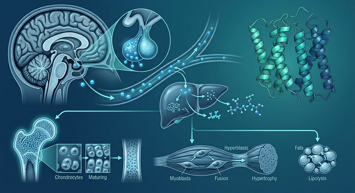
To understand why ipamorelin and CJC-1295 are used together and what they do, you need to understand the three-signal system that controls growth hormone secretion.
Growth Hormone Releasing Hormone (GHRH): Produced in the hypothalamic arcuate nucleus, GHRH travels via the portal system to the pituitary and binds GHRH receptors on somatotroph cells — the cells that synthesize and store GH. GHRH is the primary “go” signal for GH release.
Somatostatin: Also produced in the hypothalamus, somatostatin is the primary “stop” signal for GH release. It binds somatostatin receptors on pituitary somatotrophs and actively inhibits GH secretion. The interplay between GHRH (stimulatory) and somatostatin (inhibitory) produces the characteristic pulsatile pattern of GH secretion — discrete bursts occurring 4–8 times per day in healthy adults, with the largest pulse occurring shortly after sleep onset.
Ghrelin: Produced primarily in the stomach, ghrelin (the “hunger hormone”) is an endogenous ligand for the GH secretagogue receptor (GHSR-1a) on pituitary somatotrophs. Ghrelin stimulates GH release through a mechanism partially independent of GHRH — it also suppresses somatostatin activity, reducing the inhibitory brake on GH secretion.
Why pulsatility matters: GH receptors on target tissues are more sensitive to pulsatile stimulation than to continuous exposure. Continuous HGH administration produces receptor desensitization over time, reducing efficacy. The molecular mechanism operates through two parallel pathways. First, sustained GH binding activates GHR/JAK2, which phosphorylates STAT5b; STAT5b translocates to the nucleus and drives transcription of SOCS-3 (suppressor of cytokine signalling-3). SOCS-3 contains an SH2 domain that directly binds phosphorylated JAK2, sterically blocking downstream substrate access, and a SOCS-box motif that recruits an Elongin-B/C–CUL5–RBX2 E3 ubiquitin ligase complex. This complex polyubiquitinates both JAK2 and the GHR cytoplasmic domain, targeting them for proteasomal degradation. Under continuous GH stimulation, the rate of GHR internalization and proteasomal degradation exceeds the rate of receptor resynthesis, producing a net loss of surface GHR density and signal capacity. Second, ADAM10 and ADAM17 metalloproteases cleave the GHR ectodomain at the juxtamembrane stalk, releasing a soluble GH-binding protein (GHBP) into the circulation and simultaneously eliminating the membrane-anchored receptor. Continuous GH exposure upregulates ADAM17 expression, accelerating this ectodomain-shedding pathway. The pulsatile GH release produced by secretagogues (particularly the shorter-acting without-DAC versions) exploits the kinetics of SOCS-3 protein itself: SOCS-3 has a short half-life of approximately 2–3 hours and is cleared via the same ubiquitin-proteasome system it coordinates. During the GH-free trough between secretagogue pulses, SOCS-3 protein decays, ADAM17 expression returns toward baseline, and fresh GHR mRNA is translated and trafficked to the plasma membrane — restoring surface receptor density before the next pulse arrives. This cycle of signal → SOCS-3 accumulation → GHR degradation → trough recovery → resynthesis is exactly why pulsatile secretagogue delivery preserves receptor sensitivity in a way that continuous exogenous HGH administration cannot. This is the pharmacological rationale for the secretagogue approach over direct HGH replacement in non-deficiency contexts.
The Biology of Somatopause: Why GH Declines with Age
A 1991 study by Iranmanesh and colleagues documented that both the frequency and amplitude of GH secretory bursts decline significantly with age in healthy men — and that this decline is related to both chronological age and relative adiposity (Iranmanesh et al., 1991).
The mechanisms of age-related GH decline are multiple:
- Hypothalamic GHRH neuron loss: The number of GHRH-producing neurons in the hypothalamic arcuate nucleus declines with age, reducing the “go” signal. Two converging mechanisms drive this permanent neuronal attrition. First, oxidative stress-mediated mitochondrial apoptosis: arcuate GHRH neurons are large-bodied, high-output cells that sustain continuous neuropeptide synthesis, vesicular packaging, and axonal transport to the median eminence — demands that require sustained oxidative phosphorylation. Proximity to the electron transport chain generates reactive oxygen species that progressively damage mtDNA at rates 10–20× higher than nuclear DNA (due to lack of histone protection, limited mtDNA repair capacity, and physical proximity to Complex I/III ROS production sites). Somatic mtDNA point mutations accumulate in NADH dehydrogenase (Complex I) and cytochrome bc1 (Complex III) subunit-encoding genes → Complex I/III dysfunction → impaired proton gradient → compensatory electron leakage → superoxide/H₂O₂ generation in the mitochondrial matrix → pro-apoptotic Bax releases from Bcl-2/Bcl-xL sequestration → mitochondrial outer membrane permeabilization (MOMP) → cytochrome c release → apoptosome/caspase-9/caspase-3 cascade → GHRH neuron death. Because arcuate GHRH neurons are post-mitotic and cannot be replaced from a proliferating precursor pool, each apoptotic loss is permanent — the GHRH neuron pool declines monotonically. Second, sex steroid withdrawal removes trophic survival signaling: arcuate GHRH neurons express both androgen receptor (AR) and estrogen receptor alpha (ERα), and sex steroids provide two parallel neuroprotective inputs. ERα activates a rapid non-genomic Gαi-coupled PI3K/Akt signaling arm (membrane-associated ERα cross-activates IRS-1 → PI3K → Akt Thr308/Ser473) → Akt phosphorylates BAD at Ser136 → 14-3-3 sequestration of BAD in the cytoplasm → Bcl-2/Bcl-xL remain free to block Bax-mediated MOMP, protecting neurons against the oxidative mitochondrial damage that accumulates with age. AR drives hypothalamic BDNF transcription via ARE elements in the BDNF gene promoter → BDNF/TrkB sustains axonal integrity and synaptic maintenance in GHRH projection neurons independently of the Akt/Bcl-2 pathway. Andropause and menopause simultaneously withdraw both protective inputs — removing the Akt/BAD buffering and BDNF-mediated axonal maintenance — at precisely the age when cumulative mtDNA oxidative damage has already made GHRH neurons vulnerable. The two mechanisms become mutually reinforcing: declining sex steroids remove the survival signaling that was compensating for accumulated mitochondrial damage, accelerating the rate of GHRH neuron apoptosis beyond the monotonic linear rate driven by oxidative stress alone
- Increased somatostatinergic tone: Aging shifts the balance toward greater somatostatin activity through a specific hormonal mechanism: the age-related decline in sex steroids (testosterone in men, estradiol in postmenopausal women) removes a constitutive inhibitory input on periventricular nucleus (PVN) somatostatin neurons. Androgen receptor (AR) and estrogen receptor alpha (ERα) are expressed on hypothalamic PVN somatostatin neurons and function as transcriptional repressors at the somatostatin gene (SST) promoter: in young adults, liganded AR/ERα bound to androgen/estrogen response elements in the SST promoter recruit NCoR/HDAC complexes → reduced SST mRNA output per neuron → lower somatostatin release into hypophyseal portal blood. In the hypothalamus, testosterone is locally converted to estradiol by aromatase (CYP19A1) expressed in arcuate and PVN neurons, meaning much of testosterone’s central GH-axis effect is ERα-mediated. As andropause and menopause reduce circulating sex steroid levels, AR/ERα-mediated SST gene repression is withdrawn → PVN somatostatin neurons become constitutively more active → increased portal somatostatin → SSTR2/SSTR5/Gαi/adenylyl cyclase inhibition on pituitary somatotrophs → lower basal cAMP → reduced amplitude of each GH secretory pulse in response to GHRH stimulation (Giustina & Veldhuis, 1998, PMID:9733736). This sex-steroid-withdrawal-driven somatostatin increase is one reason why ipamorelin’s somatostatin-suppression arm contributes disproportionate benefit in older patients: it directly counteracts an age-amplified inhibitory tone that CJC-1295’s GHRH-R stimulation alone cannot overcome
- Adiposity amplification: Visceral adipose tissue releases free fatty acids that suppress GH pulsatility; age-related changes in body composition compound the age-related central changes
- Reduced pituitary somatotroph responsiveness: The pituitary’s capacity to respond to GHRH stimulation also declines with age — and the cellular mechanisms are now well characterized. First, GHRH receptor (GHRH-R) surface expression per somatotroph falls with age: chronic exposure to the elevated somatostatin tone described above drives GHRH-R gene transcription downward (somatostatin-mediated Gαi/cAMP suppression reduces CRE-driven GHRH-R mRNA output), and the resulting lower receptor density on each somatotroph directly limits the maximal cAMP response that any GHRH pulse can generate. Second, Gs protein expression and coupling efficiency decline in aging pituitary tissue: age-related oxidative stress produces Gs α-subunit carbonylation (particularly at Met227 and Cys254) that reduces GTP-binding affinity and the rate of GDP→GTP exchange — the molecular switch that activates adenylyl cyclase. A reduction in Gs coupling efficiency means that even when GHRH-R is occupied by ligand, the downstream cAMP signal is attenuated. Third, the L-type Ca²⁺ channel (Cav1.2/Cav1.3) phosphorylation cascade that converts cAMP into GH granule exocytosis is also compromised: protein kinase A (PKA) phosphorylation of the Cav1.2 α1C subunit at Ser1928 is a critical amplification step — phospho-Ser1928 increases single-channel open probability threefold, producing the cytosolic Ca²⁺ transient required to trigger SNARE-complex–mediated fusion of secretory granules with the plasma membrane. In aged somatotrophs, reduced cAMP amplitude translates to less PKA activation → less Ser1928 phosphorylation → smaller Ca²⁺ transients → fewer GH granules released per GHRH stimulus. These three intrinsic somatotroph defects — reduced GHRH-R density, impaired Gs coupling, and blunted PKA/Cav1.2/Ca²⁺ exocytotic gain — compound the hypothalamic changes above, explaining why ipamorelin’s ghrelin-receptor (GHSR-1a) arm, which uses a distinct Gαq/phospholipase C/IP₃/intracellular Ca²⁺ release pathway rather than the Gs/cAMP pathway, can partially rescue GH secretion even when the GHRH-R/Gs/adenylyl cyclase axis has been attenuated by age
The aggregate effect is a substantial reduction in 24-hour GH secretion and downstream IGF-1 levels with age — a pattern called somatopause. By age 70, most adults secrete approximately 25% of the GH they secreted at age 25.
The treatment controversy: Whether treating age-related GH decline is beneficial or harmful in healthy aging adults is actively debated. Proponents argue that IGF-1 normalization improves body composition, metabolic function, and quality of life in symptomatic older adults. Critics note that IGF-1 is a mitogen — it promotes cell proliferation — and epidemiological data links higher IGF-1 levels to increased cancer risk (Renehan et al., 2004). The clinical consensus is that treating documented GH deficiency (confirmed by stimulation testing) in symptomatic adults is evidence-supported, while treating “somatopause” in otherwise healthy adults without documented deficiency is a judgment call requiring individualized risk-benefit assessment.
What Are Growth Hormone Secretagogues?
Ipamorelin and CJC-1295 belong to a class of compounds called growth hormone secretagogues (GHSs) — molecules that stimulate the pituitary gland to produce and release more growth hormone. They are distinct from synthetic human growth hormone (HGH) itself.
Secretagogues stimulate your own pituitary to produce GH using the body’s existing feedback mechanisms, rather than bypassing them with exogenous hormone. The implication: secretagogues produce more physiologically regulated GH pulses than direct HGH injection, are associated with lower risk of supraphysiologic GH levels, and work via a fundamentally different mechanism than HGH replacement therapy.
Ipamorelin: Mechanism and Profile
Ipamorelin is a synthetic pentapeptide (5 amino acids: His-Aib-D-2Nal-D-Phe-Lys-NH2) that acts as a selective growth hormone secretagogue receptor (GHSR) agonist. It was developed in the 1990s by Novo Nordisk as part of research into GH deficiency treatments.
How it works: Ipamorelin binds to the ghrelin receptor (GHSR-1a) on pituitary somatotroph cells, triggering GH release. Critically, it also suppresses somatostatin release from the hypothalamus — dual action on both the “go” signal (GHSR activation) and the “stop” signal (somatostatin suppression).
Why ipamorelin over older GHRPs: First-generation GHRPs (GHRP-2, GHRP-6) elevated cortisol and prolactin substantially in addition to GH — a side effect profile that limited their clinical utility. Ipamorelin was specifically developed to be highly selective for the GH-releasing effect with minimal cortisol/prolactin stimulation (Raun et al., 1998). GHRP-6 also strongly stimulates ghrelin’s appetite-promoting effects; ipamorelin does not substantially increase appetite.
The molecular basis for ipamorelin’s superior selectivity lies in GHSR-1a receptor topology and the structural differences between GHRP compounds. The ghrelin receptor (GHSR-1a) is expressed across multiple cell populations that each respond to ghrelin-pathway agonism differently: pituitary somatotrophs at high density (GH release — the desired target); hypothalamic arcuate nucleus (ARC) NPY/AgRP neurons (appetite stimulation via ghrelin’s orexigenic pathway); pituitary corticotropes (ACTH → cortisol) and lactotrophs (prolactin) at lower density; and hypothalamic CRH neurons (CRH → ACTH cascade). Older GHRPs activate all these populations to varying degrees because of broader off-target interactions.
GHRP-6 and appetite: GHRP-6 contains a Phe-Trp pharmacophore that structurally resembles endogenous motilin — the gut peptide that regulates gastric motility. GHRP-6 partially activates motilin receptors (MLNR) expressed in the stomach and small intestine in addition to GHSR-1a, producing gastroprokinetic effects and contributing to the appetite stimulation that tracks the ghrelin/NPY/AgRP pathway. GHRP-2 and cortisol: At GHSR-1a agonist doses that produce strong GH release, GHRP-2 activates CRH neuron populations in the paraventricular nucleus (PVN) through direct GHSR-1a engagement — the ghrelin receptor is expressed on hypothalamic PVN CRH neurons at sufficient density to generate a meaningful Gq response when occupied by a high-efficacy agonist. GHRP-2’s linear peptide backbone (D-Ala²-D-βNal³-Ala⁴-Trp⁵-D-Phe⁶-Lys⁷-NH₂) provides the spatial contact geometry to engage GHSR-1a’s orthosteric binding pocket on CRH neurons with sufficient efficacy to activate Gq/11 → phospholipase C-β (PLC-β) → IP₃ + DAG: IP₃ binds IP₃ receptors on the ER of PVN CRH neurons → Ca²⁺ release → cytosolic Ca²⁺ spike → SNARE-complex-mediated CRH vesicle exocytosis from PVN axon terminals into the hypophyseal portal blood. Portal CRH binds CRF1R (CRH receptor type 1) on anterior pituitary corticotrophs — a Gs-coupled GPCR → adenylyl cyclase → cAMP → PKA → CREB Ser133 phosphorylation → CRE-driven transcription of POMC (pro-opiomelanocortin) and simultaneous PKA-mediated phosphorylation of vesicular release machinery → ACTH secretion into systemic circulation. Adrenal ACTH binds MC2R (ACTH receptor) on zona fasciculata cells → Gs/cAMP/PKA → StAR (steroidogenic acute regulatory protein) phosphorylation → cholesterol transport into the inner mitochondrial membrane → CYP11A1 (P450scc) → pregnenolone → progesterone → 17-hydroxyprogesterone → CYP11B1 (11β-hydroxylase) → cortisol secretion within 15–20 minutes of hypothalamic CRH release, generating the 2–3× baseline cortisol elevation observed in rodent studies. This cortisol elevation can persist for 60–90 minutes post-injection and chronically undermines GH axis responsiveness through GR/Pit-1 tethering, SOCS-3 induction, and somatostatin upregulation — the same mechanisms described in detail in the chronic stress section of this guide.
Ipamorelin’s structural specificity: Ipamorelin’s sequence — His-Aib-D-2Nal-D-Phe-Lys-NH₂ — incorporates a D-2-naphthylalanyl (D-2Nal) residue at position 3, the sterically bulkiest amino acid in the pentapeptide. This bicyclic naphthalene ring fills a hydrophobic subpocket in the GHSR-1a orthosteric binding site that is critical for pituitary somatotroph activation, but does so without providing the spatial contact geometry needed to activate the collateral receptor pathways on CRH neurons or motilin receptors that GHRP-2/GHRP-6 engage. The result is functional selectivity that is demonstrable at the assay level: at doses producing equivalent GH release to GHRP-6, ipamorelin produced ACTH and cortisol values indistinguishable from saline controls in rodent studies — documenting pharmacological selectivity, not just reduced efficacy (Raun et al., 1998, PMID:9849822). This selectivity profile — full GH efficacy with minimal HPA axis or appetite activation — is the primary reason ipamorelin displaced older GHRPs in clinical compounding protocols.
| GHRP | GH Release | Cortisol Elevation | Prolactin Elevation | Appetite Effect |
|---|---|---|---|---|
| GHRP-6 | Strong | Moderate | Moderate | Strong |
| GHRP-2 | Very strong | Moderate-high | Moderate | Moderate |
| Ipamorelin | Strong | Minimal | Minimal | Minimal |
| Hexarelin | Very strong | High | High | Moderate |
This selectivity is why ipamorelin became the dominant GHRP in clinical compounding protocols.
Key pharmacological characteristics:
- Half-life of approximately 2 hours — produces discrete GH pulses rather than sustained elevation
- Does not cause significant desensitization at standard clinical doses — the reason is the receptor recycling kinetics relative to the dosing interval. GHSR-1a desensitization proceeds via the canonical GRK/β-arrestin pathway: ipamorelin binding → GRK2 (G-protein-coupled receptor kinase 2) phosphorylates multiple Ser/Thr residues in the GHSR-1a intracellular C-terminal tail → β-arrestin-2 docks at these phosphosites → β-arrestin-2/receptor complex recruits clathrin and AP2 adaptor protein → clathrin-coated vesicle pinches off → endocytosis into early endosomes. However, unlike the degradative pathway operative for the GH receptor (GHR) under sustained HGH exposure — where SOCS-3 directs JAK2 polyubiquitination and ADAM17 metalloprotease sheds the GHR ectodomain, routing the receptor for proteasomal/lysosomal destruction — GHSR-1a preferentially exits early endosomes through the Rab11-marked slow recycling pathway back to the plasma membrane. During this endosomal transit, PP2A (protein phosphatase 2A) dephosphorylates the GRK2 phosphorylation sites on the GHSR-1a C-terminus, returning the receptor to the cell surface in a fully resensitized state capable of the next activation cycle. The critical pharmacokinetic factor is time: ipamorelin’s ~2-hour half-life means that at a once-daily injection, circulating drug is essentially undetectable within 8–10 hours (4–5 half-lives), leaving a ~14–16 hour trough before the next dose. During this trough, GHSR-1a recycling from early endosomes completes with a recycling half-time of approximately 20–40 minutes — the receptor restores full surface density within 2–4 hours of agonist clearance. By the time of the next injection (24 hours later, ~12 half-lives of ipamorelin), a complete complement of resensitized surface GHSR-1a awaits. GHSR-1a additionally has high constitutive internalization and recycling activity even without agonist (reflecting its ~50% constitutive basal activity), meaning the recycling machinery is continuously active and maintains a replenished receptor pool at rest. The contrast with GHR under daily HGH is mechanistically stark: GHR resynthesis requires de novo GHR mRNA transcription (Sp1/STAT5b-driven) and translation — a 12–24 hour process — meaning continuous HGH administration depletes surface GHR faster than it can be resynthesized via the Elongin-CUL5-SOCS-box ubiquitin ligase degradation pathway. GHSR-1a recycling on a 20–40 minute timescale is why pulsatile ipamorelin dosing avoids cumulative receptor loss
- Does not meaningfully stimulate appetite (unlike GHRP-6)
- Produces GH pulses that mimic the natural pulsatile GH release pattern
Preclinical evidence: Animal studies confirmed ipamorelin effectively stimulates GH release and, through downstream IGF-1 elevation, supports bone mineral density, lean body mass, and metabolic parameters with a clean toxicology profile at doses well above clinical ranges (Raun et al., 1998).
Human clinical data: Ipamorelin was studied in Phase 1 and Phase 2 clinical trials by Novo Nordisk in the early 2000s. The GH deficiency development program was discontinued not due to safety concerns but due to competitive market considerations. Phase 2 data showed effective GH stimulation in healthy volunteers, but this research was never published in full peer-reviewed form — a significant limitation on the human evidence base.
CJC-1295: Mechanism and Profile
CJC-1295 is a synthetic analog of growth hormone releasing hormone (GHRH) — the hypothalamic hormone that naturally signals the pituitary to produce and release GH. It is structurally related to sermorelin but modified for substantially longer duration.
How it works: CJC-1295 binds to GHRH receptors on pituitary somatotroph cells, stimulating GH synthesis and release. The original CJC-1295 was engineered with a Drug Affinity Complex (DAC) — a maleimide-based reactive pharmacophore attached to the GHRH peptide backbone via a lysine side-chain linker. The maleimide undergoes thiol-Michael addition with the free thiol of Cys34, albumin’s single unpaired cysteine residue in subdomain IIA: the maleimide double bond acts as a Michael acceptor, and the Cys34 thiolate (pKa ~7.9, reactive at physiological pH) attacks it nucleophilically → forming an irreversible thioether bond. The peptide-albumin conjugate is now effectively 66.5 kDa — well above the ~8 kDa renal glomerular filtration cutoff — preventing urinary clearance. Albumin additionally evades endosomal degradation through FcRn (neonatal Fc receptor)-mediated transcytosis: within endosomes at pH 6.0, FcRn binds albumin at its subdomain III/IIA interface → the complex undergoes retrograde transcytosis to the extracellular surface → at pH 7.4 affinity drops and albumin is released back into plasma. This FcRn recycling cycle sustains albumin’s 19–21 day plasma half-life; the covalently bound CJC-1295 DAC conjugate cycles with it, achieving a functional t½ of 6–8 days for the peptide portion. The without-DAC version (Mod GRF 1-29) lacks the maleimide pharmacophore; instead, four strategic amino acid substitutions resist enzymatic degradation — most critically at position 2, where native Ala² is replaced by Aib (α-aminoisobutyric acid, α-methylalanine): the Cα-methyl group creates steric bulk that prevents DPP-IV (dipeptidyl peptidase-4) from cleaving the His¹-Aib² bond (DPP-IV’s S1 pocket accommodates small aliphatic side chains but cannot engage the geminal-dimethyl Cα geometry of Aib), extending t½ from the ~7 minutes of native GHRH(1-29) to ~30 minutes — sufficient for pulsatile injection protocols while preserving discrete GH pulse architecture.
Two versions in clinical use:
| Version | Half-Life | Injection Frequency | GH Profile | Common Use |
|---|---|---|---|---|
| CJC-1295 with DAC | 6–8 days | Weekly | Sustained elevation; blunted pulsatility | Some practitioners for convenience |
| CJC-1295 without DAC (Mod GRF 1-29) | 30 minutes | Daily, paired with ipamorelin | Pulsatile; mirrors natural GHRH bursts | Most clinical protocols |
The without-DAC version (Mod GRF 1-29) is preferred for pulsatility preservation. CJC-1295 with DAC produces more continuous GH elevation — beneficial for some applications but potentially causing tachyphylaxis through a specific receptor desensitization sequence: the 6–8 day DAC-albumin half-life means GHRH-R experiences sustained agonist occupancy without a recovery trough → GRK2/GRK3 (G-protein-coupled receptor kinases) continuously phosphorylate multiple Ser/Thr residues on the GHRH-R cytoplasmic C-terminal tail → β-arrestin-2 docking → clathrin/AP2-mediated endocytosis; unlike GHSR-1a (described above), which exits early endosomes via the Rab11/PP2A resensitization pathway during agonist-free troughs, GHRH-R under persistent DAC agonism cannot achieve the agonist-dissociation required for PP2A-mediated GRK2 phosphosite dephosphorylation — instead routing to lysosomal degradation → surface GHRH-R density falls as degradation outpaces de novo receptor resynthesis (a 12–24 hour transcription/translation process), progressively attenuating the Gs/cAMP signal per unit dose. This is less of a concern with the pulsatile approach: Mod GRF 1-29’s ~30-minute half-life creates ~14–16 hour agonist-free troughs that permit GHRH-R endosomal recycling and full resensitization before each injection.
Published human clinical data: A Phase 1/2 clinical trial of CJC-1295 with DAC published in the Journal of Clinical Endocrinology & Metabolism (2006) enrolled 65 healthy adults aged 21–61. The study showed dose-dependent increases in plasma GH levels (2–10 fold above baseline) and IGF-1 elevations persisting up to 28 days after a single injection. No serious adverse events were reported; common side effects were flushing, headache, and transient dizziness — consistent with vasodilation from GH release (Teichman et al., 2006). This is the most rigorous published human clinical data for any GHRH analog in this class.
Why They Are Almost Always Used Together
Ipamorelin and CJC-1295 (or Mod GRF 1-29) work on two complementary receptor systems — the GHSR (ghrelin receptor) and the GHRH receptor respectively. Using them together produces a synergistic effect on GH release substantially larger than either compound alone.
The physiological logic mirrors the natural GH release system: GHRH stimulates the GHRH receptor (CJC-1295 mimics this), while ghrelin stimulates GHSR and suppresses somatostatin (ipamorelin mimics this). Combined, they replicate both stimulatory inputs simultaneously while reducing the inhibitory somatostatin signal.
Studies in animal models confirmed the synergistic GH response of combined GHRP + GHRH analog administration (Alba et al., 2005). Most clinical protocols for ipamorelin use it paired with Mod GRF 1-29 precisely because of this synergy and the pulsatility it preserves.
The molecular basis of synergy: two independent second-messenger cascades
The pharmacological synergy between ipamorelin and CJC-1295 is not merely “two drugs hitting different receptors.” It reflects two completely distinct intracellular signal transduction pathways that both converge on the same final step — raising intracellular calcium to drive GH granule exocytosis. Understanding the distinction explains why the combination produces GH pulses 3–5× larger than either compound alone.
CJC-1295 → GHRH-R → Gs/cAMP/PKA pathway: The GHRH receptor is a Class B secretin-family Gs-coupled GPCR. CJC-1295 binding activates Gs → adenylyl cyclase → cAMP → protein kinase A (PKA). PKA then phosphorylates L-type voltage-gated Ca²⁺ channels in the somatotroph plasma membrane → Ca²⁺ influx from extracellular space → GH granule-membrane fusion → exocytosis. Simultaneously, PKA → phospho-CREB → CRE in the GH gene promoter → new GH mRNA synthesis, replenishing the secretory pool over hours.
Ipamorelin → GHSR-1a → Gq/PLC/IP₃+DAG pathway: The ghrelin receptor (GHSR-1a) is a Gq/11-coupled GPCR — an entirely different G-protein class. Ipamorelin binding activates Gq → phospholipase C-β (PLC-β) → cleaves membrane PIP₂ into two second messengers simultaneously: (1) IP₃ binds IP₃ receptors on the endoplasmic reticulum (ER) → releases Ca²⁺ from intracellular stores → rapid cytosolic Ca²⁺ spike → calmodulin/CaM kinase II activation → GH granule exocytosis; (2) DAG remains in the plasma membrane → recruits and activates protein kinase C (PKC) → PKC phosphorylates L-type Ca²⁺ channel subunits → sustained Ca²⁺ entry → sustained GH secretion wave. Unlike the GHRH-R pathway which relies on extracellular Ca²⁺ influx, ipamorelin’s IP₃ arm mobilizes ER Ca²⁺ stores — a rapid and independent calcium source (Smith RG, 2005, PMID: 16183911).
Why the combination is synergistic, not additive: cAMP/PKA and IP₃/PKC operate independently — neither pathway’s activity is rate-limited by the other. When both are activated simultaneously, two independent mechanisms elevate intracellular Ca²⁺ in the same somatotroph: extracellular Ca²⁺ influx (via PKA-phosphorylated channels from CJC-1295) plus ER Ca²⁺ store release (via IP₃ from ipamorelin). The combined Ca²⁺ signal exceeds what either pathway achieves alone, driving larger GH granule exocytosis from the same cell. This mechanistic independence — rather than parallel pathways competing for the same limiting signaling molecule — is what produces true pharmacological synergy rather than simply additive effects.
The somatostatin suppression arm adds a third dimension: Ipamorelin’s GHSR-1a activation reduces somatostatin release from the hypothalamus — and the mechanism is indirect, operating through an arcuate nucleus relay rather than direct receptor inhibition of somatostatin neurons. GHSR-1a is expressed at high density on arcuate nucleus NPY/AgRP neurons — the same neurons that mediate ghrelin’s orexigenic signal. Ipamorelin binding activates GHSR-1a on these arcuate neurons → Gq/PLC-β → IP₃ + DAG → Ca²⁺ release from ER stores → arcuate neurons release NPY (neuropeptide Y) from axonal projections to the periventricular nucleus (PVN). Released NPY binds Y1R (NPY receptor 1, a Gi-coupled GPCR) on PVN somatostatin neurons → Y1R/Gαi → adenylyl cyclase inhibition → reduced cAMP → reduced PKA-driven Ca²⁺-dependent somatostatin vesicle fusion → less somatostatin into the hypothalamo-hypophyseal portal blood. Simultaneously, AgRP co-released from the same arcuate neurons acts as an inverse agonist at MC3R and MC4R expressed on hypothalamic PVN neurons, blocking constitutive Gs activity → additional reduction in cAMP and somatostatin output from those cells. The reason ipamorelin suppresses somatostatin without strongly stimulating appetite is that its D-2Nal structural selectivity produces lower intrinsic efficacy at arcuate NPY/AgRP neurons than at pituitary somatotrophs — sufficient NPY/Y1R activation to attenuate PVN somatostatin output, but below the threshold required to drive the strong orexigenic NPY signaling in the lateral hypothalamus and nucleus accumbens shell that constitutes meaningful appetite stimulation — a partial and selective arcuate activation that is the molecular basis for the somatostatin brake removal without the appetite drive that full ghrelin-pathway activation produces. Less somatostatin means less Gi-mediated inhibition of adenylyl cyclase in the somatotroph — allowing CJC-1295’s cAMP to accumulate to higher levels than it would if somatostatin were unopposed. Ipamorelin thus removes the brake while CJC-1295 presses the accelerator, and ipamorelin simultaneously adds its own independent Ca²⁺ mobilization signal.
IGF-1: The Downstream Effect That Drives Clinical Outcomes
GH itself has a short serum half-life (20–30 minutes). Most of GH’s biological effects are mediated through IGF-1 (insulin-like growth factor 1), produced primarily by the liver in response to GH signaling. IGF-1 has a half-life of 12–15 hours and is the clinically measurable biomarker of GH axis activity.
What IGF-1 does in target tissues:
Skeletal muscle: IGF-1 binds the IGF-1 receptor (IGF-1R) and activates the PI3K/Akt/mTOR pathway — the primary anabolic signaling cascade for muscle protein synthesis. Elevated IGF-1 promotes satellite cell (muscle stem cell) activation, nitrogen retention, and net muscle protein accretion. This is the mechanistic basis for the body composition claims associated with GH secretagogue therapy.
At the receptor level, IGF-1R engagement on skeletal myofibers and satellite cells recruits insulin receptor substrate-1 (IRS-1), which is tyrosine-phosphorylated at multiple Tyr residues by the activated IGF-1R kinase domain. Phospho-IRS-1 recruits the p85 regulatory subunit of PI3K, activating the p110 catalytic subunit to generate PIP3 at the inner leaflet of the plasma membrane. PIP3 recruits PDK1, which phosphorylates Akt at Thr308; mTORC2 simultaneously phosphorylates Akt at Ser473, achieving full kinase activation. Two parallel anabolic arms diverge from Akt. In the first, Akt phosphorylates TSC2 (tuberin) at Thr1462, inhibiting the TSC1-TSC2 GAP complex and allowing RHEB to remain GTP-loaded and mTORC1-activating. Active mTORC1 phosphorylates S6K1 at Thr389 — activating ribosomal protein S6 and eIF4B for 40S ribosome recruitment — and hyperphosphorylates 4E-BP1 at Thr37/Thr46/Ser65, releasing sequestered eIF4E to join the eIF4F cap-binding complex. The resulting burst of cap-dependent translation preferentially drives synthesis of large, abundant structural mRNAs: myosin heavy chain isoforms (MYH1/2/4/7), skeletal α-actin (ACTA1), and titin — the contractile proteins that define fiber cross-sectional area. In the second arm, Akt phosphorylates FOXO3a at Ser253 and Thr32, creating 14-3-3 docking sites that sequester FOXO3a in the cytoplasm. Nuclear exclusion of FOXO3a suppresses transcription of the E3 ubiquitin ligases MuRF1 (TRIM63) and MAFbx/atrogin-1 (FBXO32), which are the primary drivers of ubiquitin-proteasome degradation of myofibrillar proteins during atrophy. IGF-1R signaling therefore simultaneously accelerates myofibrillar protein synthesis via mTORC1/eIF4F and decelerates myofibrillar protein degradation via FOXO3a cytoplasmic retention — shifting net protein balance strongly positive through two mechanistically independent routes.
For satellite cells, a parallel IRS-1/Ras/MEK/ERK1/2 arm drives entry into the Pax7+/MyoD+ proliferating myoblast state; IGF-1R/Akt/mTORC1 subsequently promotes cap-dependent translation of the myogenic regulatory factors myogenin (MYOG) and MRF4, triggering terminal differentiation and fusion of proliferating myoblasts into existing myofibers. This satellite cell arm explains why sustained IGF-1 elevation supports long-term remodeling capacity of muscle tissue beyond the acute translational response.
Bone: IGF-1 stimulates osteoblast proliferation and differentiation, increasing bone mineral density. GH-deficient adults have significantly lower bone mineral density than age-matched controls; GH replacement studies in deficient adults show consistent improvement. Whether GH secretagogue therapy in non-deficient adults produces clinically meaningful bone density improvement is less well-established.
The osteoblast differentiation mechanism is Akt-mediated stabilization of Runx2 — the master transcription factor that drives osteoblast lineage commitment and function. IGF-1 binds IGF-1R on osteoblast precursor cells → tyrosine kinase activation → IRS-1 (insulin receptor substrate-1) phosphorylation → PI3K → PIP₃ → PDK1/Akt. Active Akt phosphorylates and inhibits GSK3β (glycogen synthase kinase 3β) at Ser9. Uninhibited GSK3β would phosphorylate Runx2 at Ser104 in its Runt homology domain, marking it for ubiquitin-mediated proteasomal degradation. When Akt suppresses GSK3β, Runx2 is stabilized — it accumulates in osteoblast nuclei and drives transcription of osteocalcin, bone sialoprotein (BSP), osteopontin, and collagen type I — the structural and mineralization proteins of bone matrix. Simultaneously, Akt → mTORC1 → S6K1 → ribosomal protein S6 phosphorylation increases translational capacity for the large collagen I and osteocalcin protein synthesis demands of active osteoblasts (Guntur & Rosen 2012, PMID:22923484). This Akt/GSK3β/Runx2 axis explains why GH-deficient patients have poor bone density despite adequate dietary calcium — the anabolic signal that drives Runx2 activity and osteoid matrix deposition is impaired at the receptor level, not the mineral supply level. The implication for GH secretagogue therapy is that the bone benefit requires sustained IGF-1 elevation over months rather than the acute metabolic effects measured at early timepoints.
Adipose tissue: IGF-1 promotes adipocyte differentiation but GH itself has a lipolytic (fat-mobilizing) effect that tends to dominate in the context of GH elevation — and the mechanism of GH-driven lipolysis is important to understand, because it operates through two parallel arms with different kinetics. In the genomic (slower) arm: GH binds GHR on adipocytes → GHR/JAK2 transphosphorylation → STAT5b Tyr694 phosphorylation → STAT5b nuclear translocation → STAT5-responsive elements in the promoter of HSL (hormone-sensitive lipase, also called LIPE) gene → increased HSL mRNA and protein synthesis over 4–8 hours. In the non-genomic (acute) arm: GH → GHR/JAK2 → IRS-1 → PI3K → Akt → but in adipocytes, unlike muscle, this Akt signal does NOT drive anabolism; instead, GH simultaneously activates a signaling branch that promotes phosphodiesterase (PDE) inhibition → cAMP accumulates in the adipocyte. The elevated cAMP activates PKA → PKA phosphorylates HSL at Ser563 and Ser660 (activating phosphorylation sites), and phosphorylates PLIN1 (perilipin-1) at Ser517, which gates access to the lipid droplet surface. Phospho-perilipin displaces CGI-58 (ABHD5) → CGI-58 is now free to activate ATGL (adipose triglyceride lipase) → ATGL initiates triglyceride hydrolysis (producing diacylglycerol), while active HSL completes the cascade (diacylglycerol → monoacylglycerol → glycerol) → net release of non-esterified fatty acids (NEFAs) and glycerol into circulation (PMID: 17409479). GH also reduces insulin sensitivity specifically in adipocytes (GHR/JAK2 → IRS-1 Ser307 phosphorylation → attenuated PI3K response to insulin → less insulin-driven re-esterification of fatty acids back into triglyceride) — ensuring that NEFAs released by lipolysis stay in circulation as fuel rather than being recaptured. The net body composition effect of GH secretagogue therapy — less visceral fat, more lean mass — therefore reflects two simultaneously operating mechanisms: GH’s direct adipocyte lipolysis program (genomic + acute PKA/HSL arms) and IGF-1’s muscle protein anabolic program through the PI3K/Akt/mTOR axis.
Nervous system: IGF-1 crosses the blood-brain barrier and has documented roles in hippocampal neurogenesis, BDNF expression, and cognitive function — with a more specific molecular basis than is usually described. IGF-1 in the peripheral circulation reaches the CNS via LRP1 (low-density lipoprotein receptor-related protein 1), a multiligand endocytic receptor expressed on brain capillary endothelial cells and the choroid plexus epithelium that mediates transcytosis of IGF-1 across the BBB into the brain parenchyma. Once in the hippocampus, IGF-1 binds IGF-1R (IGF-1 receptor) on CA1/CA3 pyramidal neurons and on neural progenitor cells (NPCs) in the dentate gyrus subgranular zone (SGZ) — the primary neurogenic niche in the adult brain. IGF-1R tyrosine kinase activation proceeds through the same IRS-1/PI3K/PIP₃/PDK1/Akt cascade as in muscle and bone, but with distinct downstream targets: Akt → phospho-Ser9-GSK-3β in hippocampal neurons reduces tau phosphorylation at the same pathological sites (Ser202, Thr231, Ser396) targeted by GLP-1R/PKA in Parkinson’s neuroprotection — meaning IGF-1’s hippocampal neuroprotection operates through direct tau stabilization as well as through the Runx2-independent Akt/GSK-3β axis relevant to Alzheimer’s pathology. A second convergent arm: IGF-1R/IRS-1 → MAPK/ERK1/2 → RSK2 → CREB Ser133 phosphorylation in hippocampal pyramidal neurons → CREB occupies CRE (cAMP response elements) in the BDNF gene promoter IV (the activity-dependent promoter that drives most cortical and hippocampal BDNF transcription) → increased BDNF mRNA synthesis. Released BDNF/TrkB then drives adult neurogenesis in the SGZ: NPC proliferation via TrkB/PLCγ/IP₃/Ca²⁺ → CaMKIV → CREB, and NPC survival via TrkB/PI3K/Akt/Bcl-2 upregulation (Trejo et al., 2001, PMID:11493516). This IGF-1→ERK→CREB→BDNF→TrkB→neurogenesis cascade is part of the mechanistic explanation for why exercise-stimulated peripheral IGF-1 elevation correlates with hippocampal volume preservation in aging adults — and why GH secretagogue therapy that sustains IGF-1 in the age-matched normal range is mechanistically coherent (though not RCT-proven) as a contributor to cognitive aging trajectories.
Sleep Architecture and GH Secretion: The Bedtime Protocol Explained
The recommendation to inject ipamorelin/CJC-1295 at bedtime is not simply about convenience. It reflects a fundamental aspect of how the GH axis operates — and understanding it helps patients optimize their protocol.
The slow-wave sleep / GH pulse relationship
Growth hormone secretion in humans is profoundly tied to slow-wave sleep (SWS), the deepest stages of non-REM sleep (N3 stage, also called delta sleep). In healthy young adults:
- The first SWS period, occurring 60–90 minutes after sleep onset, triggers the largest single GH pulse of the 24-hour period
- This nocturnal pulse can account for 70–80% of total daily GH secretion in young adults
- Subsequent sleep cycles produce smaller pulses that collectively make up most of the remaining daily GH secretion
The mechanism is specific: SWS generates a hypothalamic GHRH surge that is superimposed on low somatostatin tone (somatostatin activity is physiologically lowest during early sleep). The combination — maximal GHRH drive + minimal inhibitory somatostatin — produces the large nocturnal GH peak that drives much of the body’s nighttime anabolic activity: muscle protein synthesis, fat mobilization, immune cell proliferation, and connective tissue repair.
How aging disrupts this system
SWS declines dramatically with age. Young adults spend approximately 20–25% of total sleep time in SWS. By age 60, most adults average less than 5% SWS; many have essentially no measurable SWS. The specific relationship between SWS loss and GH decline with aging is not coincidental — they are mechanistically linked.
The cellular mechanism behind this age-related SWS loss is now partially understood and directly relevant to why GH secretagogue efficacy degrades with age independently of pituitary function. Cortical slow oscillations — the 0.5–1 Hz signature of SWS — are generated by layer V/VI pyramidal neurons in the medial prefrontal cortex (mPFC) cycling between synchronized “up states” (membrane depolarization, action potential generation) and “down states” (hyperpolarization, silence). This alternation requires precise coordination by parvalbumin-positive (PV+) GABAergic interneurons, which provide the inhibitory timing signals that synchronize large cortical networks into coherent slow oscillations. PV+ interneurons are fast-spiking cells with exceptionally high metabolic demands — they depend on dense mitochondria and efficient oxidative phosphorylation to sustain firing rates up to 600 Hz. Precisely because of this metabolic intensity, PV+ interneurons are selectively vulnerable to aging-related mitochondrial dysfunction and reactive oxygen species accumulation. As mtDNA mutations accumulate and mitochondrial membrane potential declines in aged neurons, PV+ interneuron density decreases in the prefrontal cortex — reducing the synchronizing inhibition that generates slow oscillation coherence (PMID:22037514). A second mechanism operates through the suprachiasmatic nucleus (SCN): the SCN circadian pacemaker progressively weakens with aging, reducing the amplitude of the nocturnal circadian signal (shallower core body temperature dip, attenuated melatonin peak) that consolidates early-night sleep when the largest GH pulse is generated. Finally, the ventrolateral preoptic nucleus (VLPO) — the hypothalamic sleep-promoting nucleus containing galanin/GABA neurons that inhibit wake-promoting arousal systems (locus coeruleus norepinephrine neurons, dorsal raphe serotonin neurons, tuberomammillary nucleus histamine neurons) — undergoes selective neurodegeneration with aging, its progressive cell loss reducing inhibitory drive through a dual co-transmitter mechanism that explains why VLPO attrition is so disruptive: VLPO neurons simultaneously release GABA (binding GABA-A receptors, ionotropic Cl⁻ channels, on LC/raphe/TMN neuron soma and dendrites → Cl⁻ influx → rapid membrane hyperpolarization → action potential threshold raised → NE, 5-HT, and histamine release suppressed within milliseconds) and galanin (binding GALR1, a Gi/o-coupled GPCR, on the same arousal neurons → Gαi/Gβγ → GIRK (Kir3.1/Kir3.2) inwardly rectifying K⁺ channel opening via direct Gβγ-channel subunit binding → sustained K⁺ efflux → membrane hyperpolarization maintained for minutes beyond the Cl⁻ transient); the GABA-A component is fast but desensitizes rapidly under tonic release; the GALR1/GIRK component is slower-onset but does not desensitize at the same rate, sustaining inhibition through the critical hours of early-night SWS; when VLPO neurons degenerate, both the fast GABA-A and the sustained GALR1/GIRK inhibitory inputs to LC, raphe, and TMN are simultaneously withdrawn → these arousal nuclei are disinhibited → NE, 5-HT, and histamine release fragment early-night SWS at precisely the window when the nocturnal GHRH-driven GH pulse should be occurring. These three convergent mechanisms — PV+ interneuron loss impairing slow oscillation generation, SCN weakening reducing circadian sleep consolidation, and VLPO degeneration allowing arousal breakthrough — together explain why the natural nocturnal GHRH surge fails to produce an adequate GH pulse in older adults: the SWS architecture that gates the GHRH surge is structurally degraded before the GH axis itself has failed. The clinical implication is that sleep quality restoration (CPAP for OSA, sleep hygiene optimization, addressing blue light and alcohol) improves secretagogue response by rebuilding the SWS substrate the bedtime injection is designed to amplify.
Van Cauter and colleagues documented this relationship in a landmark 2000 study: the decline in GH secretion with aging is tightly correlated with the simultaneous decline in SWS (Van Cauter et al., 2000, PMID: 10768168). Aging men who retained more SWS retained more GH secretion. The GH axis wasn’t simply failing at the hypothalamic level — the sleep-GH relationship was weakening together.
The glymphatic connection: SWS is also when the brain’s glymphatic clearance system operates most efficiently — clearing metabolic waste products including amyloid-beta and tau. GH secretagogues that improve the quantity and depth of SWS may thus have cognitive benefits extending beyond the GH axis through this glymphatic pathway. The molecular sequence is specific: glymphatic clearance depends on AQP4 (aquaporin-4) water channels densely clustered at astrocyte endfeet — polarized astrocytic extensions that sheathe cerebral blood vessels. During SWS, synchronized neuronal “down states” (the hyperpolarization phase of the slow cortical oscillation generated by PV+ interneuron-coordinated layer V pyramidal neuron cycling) reduce neuronal metabolic demand, lowering local interstitial osmolarity; simultaneously, low-frequency, high-amplitude arterial pulsations during SWS rhythmically expand and contract perivascular spaces. These two forces combine: CSF enters para-arterial spaces between vessel walls and AQP4-rich astrocyte endfeet → AQP4 channels mediate bulk water movement down the osmotic gradient from the parenchymal interstitium into perivascular space → convective flow entrains amyloid-beta (Aβ40/42), hyperphosphorylated tau, and other metabolic waste along para-arteriolar paths → perivenous drainage → meningeal and cervical lymphatic clearance. SWS disruption — including the VLPO-degeneration/PV+ interneuron loss mechanisms described above — reduces this convective clearance by approximately 60% compared to consolidated SWS, directly linking age-related sleep fragmentation to Aβ and tau accumulation. GH secretagogues add a second, mechanistically distinct contribution beyond the SWS-architecture arm: GH receptors (GHR) are expressed on cortical and hippocampal astrocytes. Nocturnal GH pulse → GHR/JAK2/STAT5b in astrocytes → STAT5b-responsive elements in the AQP4 gene promoter → increased AQP4 mRNA transcription. A parallel IGF-1 arm: GH → hepatic IGF-1 → IGF-1R on astrocytes → PI3K/Akt/mTOR → AQP4 protein synthesis and polarized trafficking to astrocyte endfeet — precisely the location where the perivascular hydraulic conductance is determined. Higher endfeet AQP4 density increases the conductivity of the astrocyte endfeet layer, allowing the SWS-driven perivascular pressure gradient to move more water and entrained solutes per oscillation cycle. GH secretagogues therefore contribute to glymphatic clearance through two separable mechanisms: amplifying the SWS architecture that provides the fluid-dynamic driving force, and elevating GH/IGF-1 to increase the AQP4 channel capacity that executes it — a convergence that explains why age-related somatopause, SWS decline, and Aβ accumulation track together so closely in the aging brain.
Why bedtime injection amplifies the natural pulse
A bedtime ipamorelin/CJC-1295 injection — administered 30–60 minutes before sleep onset, on an empty stomach — is designed to amplify the naturally occurring nocturnal GHRH pulse:
- CJC-1295 (Mod GRF 1-29) adds GHRH receptor stimulation, enhancing the pituitary’s GH-producing response
- Ipamorelin suppresses somatostatin and activates GHSR, removing the inhibitory brake while adding ghrelin-pathway drive
- Together, they amplify a pulse that was already primed to occur by the sleep architecture
This is additive pharmacology, not replacement. The secretagogues aren’t forcing an artificial GH pulse at an arbitrary time; they’re enlarging a pulse the body was already generating. This is why pulsatility is preserved and why the physiological feedback mechanisms remain functional — the surge occurs, triggers negative feedback, and returns to baseline, mimicking (but amplifying) normal GH physiology.
Factors that impair the sleep-GH axis
Several common conditions disrupt the sleep-GH relationship and reduce the effectiveness of bedtime secretagogue injection:
Obstructive sleep apnea (OSA): Apnea episodes cause repeated SWS disruptions. Patients with untreated OSA have severely blunted nocturnal GH pulses. Treating OSA (CPAP or positional therapy) before optimizing GH axis supplementation is logical — restoring SWS structure significantly improves the natural GH pulse that secretagogues are designed to amplify.
Alcohol: Even moderate evening alcohol use suppresses SWS in the early night sleep period — precisely when the largest GH pulse is generated. Patients who drink regularly in the evenings will have impaired GH secretagogue responses. This is a modifiable factor worth addressing.
Blue light/late-night screens: Blue light suppresses melatonin, delaying sleep onset and reducing SWS in the first sleep cycles. Optimizing sleep hygiene to protect early-night SWS is a practical complement to secretagogue protocols.
Chronic stress: HPA axis activation (elevated cortisol) suppresses GH secretion. Patients in chronic stress states — elevated evening cortisol, elevated CRH — will have attenuated secretagogue responses. This is another reason why GH secretagogue protocols should be evaluated in the context of overall HPA axis function.
Molecular mechanism of cortisol-mediated GH suppression: Cortisol acts through three distinct mechanisms that converge to blunt CJC-1295 and ipamorelin efficacy:
(1) Pituitary somatotroph GH transcription suppression: Cortisol/GR (glucocorticoid receptor) complex translocates to the somatotroph nucleus → GR physically tethers via protein–protein interaction to Pit-1 (POU1F1), the pituitary-specific transcription factor that drives GH gene expression by binding PISCES elements (pituitary-specific elements) in the GH gene promoter. The structural basis: GR’s DNA-binding domain (DBD) D-box region contacts the POU homeodomain (POUhd) α-helix 3 of Pit-1 — the same recognition helix that normally contacts PISCES DNA major-groove base pairs to drive GH transcription. This D-box/POUhd3 protein–protein contact sequesters the Pit-1 recognition helix away from PISCES DNA while keeping the GR/Pit-1 complex anchored at the GH promoter locus, explaining why the repression is promoter-specific rather than global. Simultaneously, the cortisol-bound GR ligand-binding domain (LBD) adopts a repressive conformation in which helix 12 is retracted (rather than positioned in the agonist “AF-2” cleft): this retracted-H12 surface exposes the CoRNR-box binding groove on the GR LBD, which recruits NCoR/SMRT via their RID1/RID2 CoRNR-box motifs (LXXI/HIXXXI/L) → NCoR/SMRT scaffold recruits Sin3A → HDAC3 to the GH upstream regulatory region → HDAC3 deacetylates H3K9ac and H4K8ac at the GH gene locus → chromatin compaction → reduced RNA Pol II elongation at GH mRNA → reduced GH mRNA synthesis. This means the somatotroph granule pool replenishment rate falls even when CJC-1295 drives GHRH-R activation — the secretion machinery is stimulated but the substrate being secreted is in shorter supply.
(2) SOCS-mediated JAK/STAT signal attenuation: The GR/glucocorticoid complex additionally drives transcription of SOCS-2 and SOCS-3 (suppressor of cytokine signaling proteins) in somatotrophs → SOCS proteins contain an SH2 domain that binds phosphotyrosine residues on activated JAK2 → competitive inhibition of JAK2 kinase activity → reduced phosphorylation of STAT5b (the primary transcription factor of the GHRH-R/Gs/cAMP downstream axis) → blunted signal amplification even when CJC-1295 occupies GHRH-R at full receptor saturation. SOCS-3 additionally targets the insulin receptor substrate adapter proteins involved in the GH receptor signaling cascade, compounding IGF-1 axis suppression downstream.
(3) Hypothalamic somatostatin upregulation: Cortisol/GR in periventricular nucleus somatostatin neurons → GR binds GRE (glucocorticoid response elements) in the somatostatin gene promoter → increased somatostatin (SST) mRNA and peptide synthesis → elevated somatostatin release into the hypothalamo-hypophyseal portal circulation → SST binds SSTR2 (the dominant pituitary somatostatin receptor subtype) on somatotrophs → SSTR2 couples to Gαi → adenylyl cyclase inhibition → reduced cAMP → reduced PKA activity → reduced L-type Ca²⁺ channel phosphorylation and reduced Ca²⁺-dependent GH granule exocytosis. Because CJC-1295 works by amplifying cAMP through GHRH-R/Gs, elevated somatostatin tone creates an opposing Gi signal that directly undermines the CJC-1295 mechanism regardless of dose.
The clinical implication: CJC-1295 + ipamorelin administered to a patient with chronic stress-driven HPA activation faces simultaneous suppression of GH gene transcription (mechanism 1), blunted GHRH-R signal amplification (mechanism 2), and active Gi-mediated cAMP antagonism (mechanism 3). Normalizing HPA axis function — through stress reduction, sleep optimization, and addressing root causes of chronically elevated cortisol — is therefore not optional optimization but a prerequisite for meaningful secretagogue response.
Practical Protocol Guidance: Timing and Administration
While specific dosing should be established by your prescribing provider, understanding the rationale behind common protocol elements helps patients use these compounds most effectively.
Fasting state matters: Insulin is an endogenous suppressor of GH secretion. A large carbohydrate or sugar intake raises insulin, which blunts the GH pulse produced by secretagogue administration. For this reason, most protocols specify:
- Inject either well before a meal (30–60 minutes before eating) or 2+ hours after the last meal/food intake
- Avoid administering with carbohydrate-rich foods
The mechanism by which postprandial glucose and insulin suppress GH operates through two converging pathways that both converge on somatostatin — making them directly antagonistic to CJC-1295’s mechanism of action:
Pathway 1 — Hypothalamic glucose-sensing via KATP channels in somatostatin neurons. Carbohydrate ingestion → rapid intestinal glucose absorption → portal hyperglycemia reaching hypothalamic periventricular nucleus (PVN) somatostatin neurons. These neurons contain ATP-sensitive potassium (KATP) channels composed of Kir6.2/SUR1 subunits — the same channel class that mediates glucose-dependent insulin secretion in pancreatic β-cells. As glucose is metabolized in somatostatin neurons, the intracellular ATP/ADP ratio rises → KATP channels close → membrane depolarization → activation of voltage-gated Ca²⁺ channels (Cav2.1/P/Q-type in hypothalamic neurons) → cytosolic Ca²⁺ spike → SNARE complex-mediated somatostatin vesicle fusion and exocytosis → elevated portal somatostatin concentration. This somatostatin reaches pituitary somatotrophs via the hypothalamo-hypophyseal portal circulation and binds SSTR2 (the dominant pituitary somatostatin receptor subtype) → SSTR2/Gαi → adenylyl cyclase inhibition → reduced cAMP → reduced PKA activation → reduced L-type Ca²⁺ channel phosphorylation → reduced GH granule exocytosis. Critically, this Gi-mediated cAMP reduction directly antagonizes CJC-1295’s mechanism: CJC-1295 works by activating GHRH-R/Gs/cAMP, and SSTR2/Gi produces the opposing signal in the same somatotroph. The two signals are pharmacologically opposed — elevated postprandial somatostatin tone literally cancels the cAMP accumulation that CJC-1295 is trying to produce.
Pathway 2 — Insulin direct pituitary effect. The postprandial insulin surge reaches pituitary somatotrophs, which express insulin receptors (InsR). InsR activation → IRS-1 tyrosine phosphorylation → PI3K → PIP₃ → Akt. In somatotrophs, Akt activates PDE3B (phosphodiesterase 3B) — a cAMP-hydrolyzing enzyme — accelerating the breakdown of cAMP even when GHRH-R/Gs is stimulating its production by CJC-1295. PDE3B belongs to the same phosphodiesterase class that is inhibited by cilostazol and milrinone; in cardiomyocytes this pathway is pro-inotropic (cardiac cAMP elevation), but in somatotrophs where the starting cAMP from CJC-1295 is modest, insulin-driven PDE3B activation creates a “futile cycle” that prevents cAMP from accumulating to the threshold required for PKA activation and GH secretion. Additionally, elevated insulin → hepatic IGF-1 production remains adequate despite the GH suppression (insulin and GH work synergistically on IGF-1 production), but the direct pituitary suppression reduces the GH pulse height that CJC-1295 is intended to amplify.
The fasting protocol’s rationale in molecular terms: 2 hours post-meal ensures portal glucose has cleared (restoring KATP channel activity to baseline → minimal somatostatin output), insulin has returned to near-basal levels (removing PDE3B stimulation in somatotrophs), and free fatty acids have stabilized to normal range. With somatostatin tone low and PDE3B activity at baseline, CJC-1295’s cAMP accumulates unimpeded and ipamorelin’s IP₃/Ca²⁺ mobilization adds its independent calcium signal — together producing the maximum GH pulse the fasted pituitary can generate.
Timing relative to sleep: The largest natural GH pulse in healthy adults occurs within the first hour of deep (slow-wave) sleep. An ipamorelin + Mod GRF 1-29 injection at bedtime (on an empty stomach) aims to amplify this natural pulse — adding secretagogue stimulation to the existing nocturnal peak. Many protocols use a bedtime injection as the primary or sole daily injection.
Typical dose ranges used clinically: Most clinical protocols use 100–300mcg ipamorelin with 100–300mcg Mod GRF 1-29 per injection. These are not FDA-approved doses — they are practitioner-derived based on the published pharmacological data and clinical experience. The IGF-1 response, not the dose, should guide optimization.
Injection frequency: Most protocols start with one injection at bedtime (once daily) and adjust based on IGF-1 response. Some protocols use twice or three-times-daily injection in younger patients or those seeking more aggressive GH stimulation; this is less common and requires more active monitoring.
What the Research Does and Does Not Support
Reasonably well-supported:
- Both compounds effectively stimulate GH and downstream IGF-1 production in humans
- CJC-1295 with DAC has published Phase 2 human data demonstrating GH and IGF-1 elevation
- Ipamorelin has a cleaner hormone selectivity profile than older GHRPs — significantly lower cortisol and prolactin elevation
- The pulsatile GH release pattern of Mod GRF + ipamorelin more closely mimics natural GH physiology than continuous HGH
Less well-established:
- Long-term clinical outcomes (body composition, bone density, muscle mass, cognition) from secretagogue protocols in healthy aging adults have not been established in rigorous long-term RCTs
- Optimal dosing regimens used in clinical practice are derived from practitioner experience and short-term trials, not long-term efficacy studies
- Whether reversing somatopause in otherwise healthy adults with age-related GH decline (without documented deficiency) produces net clinical benefit is contested
What evidence exists for body composition: The body composition data from GH secretagogue use in healthy aging adults is almost entirely from short-term studies measuring IGF-1 response rather than body composition outcomes. The assumption that elevated IGF-1 → improved body composition is mechanistically reasonable but has not been confirmed in the population most commonly using these compounds (healthy adults aged 40–70 without documented GH deficiency) in controlled trials with body composition as the primary endpoint.
How These Compare to Sermorelin
Sermorelin is an older GHRH analog (29 amino acids of the natural GHRH sequence) that the FDA approved in 1997 for GH deficiency diagnosis and treatment in children. It is often discussed alongside ipamorelin and CJC-1295.
| Sermorelin | Mod GRF 1-29 / CJC-1295 | Ipamorelin | |
|---|---|---|---|
| Mechanism | GHRH analog | GHRH analog | GHRP (ghrelin receptor) |
| Half-life | ~10 min | ~30 min / 6–8 days | ~2 hours |
| FDA status | Formerly approved (pediatric GH deficiency); compoundable | Not FDA-approved; compoundable | Not FDA-approved; compoundable |
| Human trial data | Yes (pediatric GH deficiency) | Yes (CJC-1295 Phase 2, 2006) | Limited (unpublished Novo Nordisk trials) |
| Clinical use | Adult longevity/anti-aging (off-label) | Adult longevity/anti-aging | Typically combined with GHRH analog |
Sermorelin’s advantage is its historical FDA approval record and longer clinical track record. Its limitation is a very short half-life (~10 minutes), requiring careful injection timing to catch natural GH release windows — and the molecular basis for this short half-life explains exactly why the subsequent GHRH analogs were engineered differently. Sermorelin retains the native 29-amino acid GHRH sequence including an Ala at position 2 — the penultimate residue from its free N-terminal His¹. This Ala² is the canonical DPP-IV recognition site: DPP-IV (dipeptidyl peptidase-4, CD26) is a serine protease of the prolyl oligopeptidase family whose catalytic triad (Ser630/Asp708/His740) cleaves dipeptides from N-termini carrying Pro or Ala at the P1 penultimate position. Sermorelin’s His¹-Ala² bond is cleaved by circulating plasma DPP-IV within 2–4 minutes of injection, generating GHRH(3-29) — a fragment with negligible GHRH-R binding affinity because the His¹-Ala² N-terminal sequence is required for correct insertion into the transmembrane domain orthosteric pocket of the GHRH receptor (the same two-step ECD-capture/TMD-activation architecture as GLP-1R engagement by native GLP-1). This DPP-IV inactivation mechanism is identical to how native GLP-1 is cleaved at its His⁷-Ala⁸ bond within 1–2 minutes of L-cell secretion. Additionally, sermorelin has no albumin-binding modification; at approximately 1,357 daltons, it is efficiently filtered at the renal glomerulus and further degraded by tubular brush-border peptidases. The combination of DPP-IV N-terminal cleavage (primary inactivation, within minutes) and renal clearance (secondary) produces the ~7–10 minute functional half-life. Mod GRF 1-29 (CJC-1295 without DAC) specifically engineered around this problem: the Ala² → Aib (α-aminoisobutyric acid) substitution at position 2 places a geminal-dimethyl group on the α-carbon, creating steric bulk that prevents DPP-IV’s substrate-binding pocket from accommodating the modified residue — exactly the same Aib strategy used in semaglutide to block DPP-IV cleavage of GLP-1’s His-Ala N-terminus. Three additional substitutions in Mod GRF 1-29 (Gln²⁸→Ala, Ser²⁹→Ala, and D-Ala at position 1) reduce susceptibility to C-terminal carboxypeptidases and further plasma peptidases, extending the half-life from ~7 minutes to ~30 minutes. CJC-1295 with DAC then adds the maleimide-Cys34 covalent albumin bond that borrows albumin’s FcRn recycling half-life for a 6–8 day duration. Sermorelin’s regulatory history as a 1997 FDA-approved drug — developed before the era of systematic pharmacokinetic engineering applied to peptide half-life — explains why it retained the natural GHRH N-terminus with its DPP-IV vulnerability, while the subsequently developed analogs specifically designed for clinical longevity use engineered around the same degradation mechanism. Mod GRF 1-29 offers a longer action window with the same receptor mechanism, providing more flexibility.
Regulatory Status
In the United States (as of 2026):
- Ipamorelin and CJC-1295 (including Mod GRF 1-29) are not FDA-approved for any indication
- They are eligible for compounding through 503A and 503B pharmacies under the FDA’s bulk drug substance framework — unlike BPC-157 and TB-500, which are explicitly ineligible
- The FDA’s oversight of peptide compounding has intensified since 2023 — verify the current compounding status with your prescribing physician and pharmacy, as this landscape evolves
- Sermorelin: formerly had pediatric FDA approval (Geref), discontinued by manufacturer in 2008; has been compounded through a different regulatory pathway since
Drug Interactions That Affect Response
Several medications and health states affect GH secretagogue efficacy — patients should discuss these with their prescribing provider:
Corticosteroids: Chronic corticosteroid use significantly suppresses endogenous GH secretion and blunts pituitary response to GHRH stimulation. Patients on prednisone, hydrocortisone, or other steroids will have markedly attenuated IGF-1 responses to secretagogue therapy. This is a clinical limitation worth discussing before starting.
Thyroid dysfunction: Hypothyroidism impairs GH secretion and IGF-1 production. The mechanism: Thyroid hormones — specifically T3 (triiodothyronine), the biologically active form — are required at multiple nodes of the GH axis. In pituitary somatotrophs, T3 binds the nuclear thyroid hormone receptor alpha (TRα), which is constitutively associated with thyroid hormone response elements (TREs) in the GH gene promoter as a homodimer or TRα/RXR heterodimer. In the unliganded state (as in hypothyroidism), TRα bound to TRE recruits NCoR (nuclear corepressor) and SMRT corepressor complexes → HDAC3 deacetylates histones H3K9 and H3K14 at the GH promoter → chromatin compaction → reduced GH mRNA basal transcription. When T3 is present, it binds the ligand-binding pocket of TRα → conformational change displaces corepressor complexes → recruits SRC-1/SRC-3 co-activators and CBP/p300 histone acetyltransferases → histone acetylation → chromatin opening → RNA Pol II engagement → GH gene expression. A second critical node: T3 drives transcription of Pit-1 (POU1F1) — the pituitary-specific transcription factor whose binding to PISCES elements in the GH promoter is essential for somatotroph-specific GH expression. In hypothyroidism, reduced T3 → reduced Pit-1 expression → fewer Pit-1 molecules available to occupy GH promoter PISCES elements → blunted GH transcription even when GHRH-R/Gs/cAMP/PKA signaling is fully activated by CJC-1295. The practical implication: CJC-1295 stimulates cAMP and PKA in somatotrophs, but if Pit-1 protein levels are low due to hypothyroidism, PKA-mediated CREB phosphorylation cannot drive adequate GH gene transcription because the basal transcriptional machinery at the GH promoter is insufficient. This is why thyroid status optimization before starting GH secretagogue therapy is clinically important — it’s one of the most common modifiable factors affecting secretagogue response, and fixing it restores both the Pit-1 transcriptional machinery and the T3/TRE-mediated basal GH gene accessibility that CJC-1295 needs to work.
Diabetes medications: GH is insulin-counterregulatory — and the mechanism explains why monitoring should address bidirectional glucose variability, not only the risk of elevated glucose. GH drives hepatic glucose production through a specific transcriptional program: GH binds GHR on hepatocytes → GHR dimerization → JAK2 transphosphorylation → STAT5b Tyr694 phosphorylation → STAT5b homodimerization → nuclear translocation → STAT5b binds response elements in the promoters of G6PC (glucose-6-phosphatase, which releases free glucose from glucose-6-phosphate into the portal circulation) and PCK1 (PEPCK, phosphoenolpyruvate carboxykinase 1, the rate-limiting gluconeogenic enzyme that converts oxaloacetate to phosphoenolpyruvate). Elevated G6PC and PCK1 expression increases hepatic glucose output directly into portal blood, raising fasting blood glucose independently of peripheral insulin resistance. A parallel metabolic arm amplifies this: GH-driven lipolysis (the STAT5b/HSL/ATGL mechanism described in the IGF-1 section above) raises plasma free fatty acids → intrahepatic FFA oxidation → acetyl-CoA accumulation → allosteric activation of pyruvate carboxylase (converting pyruvate to oxaloacetate) → more oxaloacetate substrate for PCK1 → amplified gluconeogenic flux.
For insulin-dependent patients, this creates a bidirectional glucose risk: if insulin dose is not adjusted downward at secretagogue initiation, the combination can produce hyperglycemia during the GH pulse (ipamorelin + CJC-1295 peak effect, roughly 1–3 hours post-injection). But if insulin is reduced and the GH pulse subsequently subsides — ipamorelin’s ~2-hour half-life means hepatic STAT5b-driven glucose output returns toward baseline within hours — residual insulin activity may exceed the lower post-trough glucose supply, producing a hypoglycemia window at 5–8 hours post-injection. Glucose monitoring at both the GH pulse peak (~2–3 hours) and the trough (~5–8 hours) during the first weeks of combined therapy is warranted, with dose adjustment protocols discussed with the prescribing physician explicitly before starting.
Obesity: Adiposity suppresses GH pulsatility through multiple mechanisms, including free fatty acid-mediated somatostatin activation. Obese patients typically have blunted IGF-1 responses to secretagogue therapy compared to lean patients — a relevant expectation-setting point.
The free fatty acid → somatostatin pathway works as follows. Visceral adipose tissue in obese individuals releases elevated non-esterified fatty acids (NEFAs) into the portal circulation. Elevated portal FFAs are detected by the hypothalamus, where they activate GPR40 (free fatty acid receptor 1, FFAR1) and GPR120 (FFAR4) expressed on hypothalamic neurons in the periventricular nucleus (PVN) and arcuate nucleus — the same regions that contain somatostatin-producing neurons. FFAR1/GPR40 couples through Gq → PLC-β → IP₃/DAG → Ca²⁺ → calmodulin-dependent release of somatostatin from periventricular PVN neurons into the hypothalamo-pituitary portal circulation. The elevated somatostatin then binds SST2R and SST5R on pituitary somatotrophs → Gi/o → adenylyl cyclase inhibition → reduced cAMP → blunted PKA activation → reduced L-type Ca²⁺ channel opening → reduced GH secretory response to any given GHRH/secretagogue stimulus (PMID:8836508). A second mechanism operates in parallel: elevated portal FFAs directly activate PKC in hepatocytes, which reduces GH receptor signaling through IRS-1 serine phosphorylation, limiting IGF-1 production even when GH secretion is adequate. This explains why obese patients often have simultaneously low IGF-1 and low GH — the suppression operates at both the hypothalamic-pituitary level (more somatostatin) and the hepatic level (impaired GHR → IGF-1 signaling). Clinically, meaningful weight loss — even 10–15% of body weight — can restore GH pulsatility by reducing portal FFA load and hypothalamic somatostatin tone, improving secretagogue response independently of any dose change.
Alcohol: Acute alcohol consumption suppresses GH secretion through three converging mechanisms that are directly relevant to GH secretagogue efficacy. GABA-A potentiation → somatostatin disinhibition: Ethanol is a positive allosteric modulator of GABA-A receptors (ionotropic Cl⁻-conducting channels), particularly those containing α1β2γ2 subunit combinations expressed throughout the hypothalamic periventricular nucleus. Ethanol binding at the transmembrane interfaces between β and α/γ subunits stabilizes the channel’s open-probability — enhanced Cl⁻ influx hyperpolarizes GABAergic interneurons that normally suppress periventricular somatostatin neuron firing. When these interneurons are hyperpolarized, they release less GABA → somatostatin neurons are disinhibited → elevated somatostatin output into the hypothalamo-pituitary portal blood → SSTR2/Gαi/adenylyl cyclase inhibition on somatotrophs → reduced cAMP → directly opposes CJC-1295’s Gs/cAMP mechanism. Cortisol elevation: Ethanol stimulates hypothalamic CRH release → anterior pituitary ACTH → adrenal cortisol secretion within 30–60 minutes of moderate alcohol intake. This acute cortisol elevation engages the same GR/SOCS-3/somatostatin axis described in the chronic stress section above — compounding the GABA-A-driven somatostatin increase with a second cortisol-mediated somatostatin amplification arm and additionally suppressing GH gene transcription via GR/Pit-1 tethering. Sleep architecture disruption: While alcohol at moderate doses shortens sleep onset latency, it reduces slow-wave sleep (SWS) in the second half of the sleep period and fragments the nocturnal SWS architecture that the bedtime secretagogue injection is designed to amplify. Even when GH secretion per pulse is not fully blocked, SWS reduction attenuates the number and amplitude of naturally occurring GHRH surges on which ipamorelin and CJC-1295 stack their pharmacological signal. Controlled studies document 50–75% blunting of the nocturnal GH pulse after moderate alcohol consumption — making evening alcohol one of the highest-impact single modifiable variables for secretagogue response, and a practical counseling point often underemphasized in clinical protocol discussions.
Side Effects and Monitoring
Commonly reported:
- Water retention (edema), particularly in hands and feet — GH-mediated fluid accumulation operates through three parallel renal mechanisms. First, direct tubular action: GH activates GHR/JAK2/STAT5b signaling in renal collecting duct principal cells, upregulating epithelial sodium channel (ENaC) subunit expression (α, β, γ) and Na⁺/K⁺-ATPase basolateral pump density — net effect is increased luminal Na⁺ reabsorption drawing osmotic water with it. Second, aldosterone amplification: GH-stimulated IGF-1 acts on adrenal zona glomerulosa cells via IGF-1R/IRS-1/PI3K/Akt → phosphorylation of StAR (steroidogenic acute regulatory protein) at Ser194/195 → increased StAR mitochondrial cholesterol transport activity → more substrate reaching CYP11A1; and Akt phosphorylates and activates SF-1 (steroidogenic factor 1, NR5A1), the nuclear receptor that binds steroidogenic factor response elements (SFREs) in the CYP11B2 (aldosterone synthase) gene promoter → increased CYP11B2 mRNA → increased aldosterone synthase protein → increased aldosterone secretion → mineralocorticoid receptor/ENaC transcriptional amplification that compounds the direct GH tubular effect. Third, ADH/AQP2 sensitization: the mild hypo-osmolality produced by Na⁺ retention triggers hypothalamic osmoreceptor-mediated ADH (vasopressin) release → V2R/Gs/cAMP/PKA → aquaporin-2 (AQP2) phosphorylation and apical membrane insertion in collecting duct principal cells → free water reabsorption completing edema formation. Fluid accumulates preferentially in hands, feet, and ankles because hydrostatic pressure at dependent and peri-articular sites favors transcapillary filtration exceeding local lymphatic return. Resolution occurs as GH receptor desensitization via GRK2/β-arrestin attenuates the tubular signal and aldosterone output normalizes — explaining why edema is worst in the first weeks and self-limits with continued use; dose reduction accelerates resolution by reducing the upstream GH stimulus driving all three limbs simultaneously
- Flushing, headache, or lightheadedness shortly after injection — this is a direct consequence of two parallel, mechanistically distinct vasodilatory signals that occur simultaneously in the first 15–30 minutes after ipamorelin + CJC-1295 administration. The first signal is GHSR-1a/Gq-driven endothelial eNOS activation: ipamorelin binds GHSR-1a on vascular endothelial cells → Gq/PLC-β → IP₃ → IP₃R-mediated Ca²⁺ release from the endoplasmic reticulum → cytosolic [Ca²⁺] rises within seconds → Ca²⁺ binds calmodulin → the Ca²⁺/CaM complex relieves the auto-inhibitory tethering between eNOS’s oxygenase domain and its reductase domain → eNOS is disinhibited → L-arginine oxidation → nitric oxide (NO) is generated → NO diffuses across the endothelial basement membrane into adjacent vascular smooth muscle → soluble guanylyl cyclase (sGC) → cGMP → PKG-I → MYPT1 (myosin phosphatase targeting subunit 1) phosphorylation → activated myosin light-chain phosphatase (MLCP) → dephosphorylation of MLC20 → VSMC relaxation → arteriolar dilation. This NO burst is acute — it peaks within minutes of endothelial Ca²⁺ elevation and precedes any systemic GH pulse. The second signal comes from the GH pulse itself, which follows within 15–30 minutes: GH binds GHR on endothelial cells → GHR/JAK2 → a non-genomic, rapid Src kinase activation → Src → PI3K/Akt → eNOS Ser1177 phosphorylation → sustained second wave of NO production that extends arteriolar dilation beyond the initial IP₃/Ca²⁺ burst. Both NO signals converge on the same cGMP/PKG/MLCP/MLC20 pathway but are generated by distinct upstream triggers — the ipamorelin GHSR-1a/Gq/Ca²⁺ arm is immediate; the GH/JAK2/Src/Akt/eNOS arm is slightly delayed and more sustained. The combined NO output produces rapid peripheral arteriolar dilation → decreased systemic vascular resistance → flushing of the face, chest, and extremities as local capillary beds dilate. The headache mechanism is identical to the nitroglycerin headache: NO-mediated dilation of meningeal and dural blood vessels → mechanical stretch of dural mechanoreceptors (C and Aδ fibers) → trigeminovascular afferent activation → pain referral through the trigeminal-cervical complex as a throbbing, pressure-type headache. The lightheadedness reflects the same peripheral vasodilation creating a transient orthostatic challenge — cardiac output has not yet compensated for the drop in peripheral resistance, and if the patient is upright, reduced cerebral perfusion pressure for 2–5 minutes produces a pre-syncopal sensation. Bedtime injection substantially reduces all three symptoms: the recumbent position eliminates the orthostatic component (cardiac output and MAP are maintained without the gravitational demand), the total vasodilatory response is the same but the patient is asleep through the peak NO phase, and the additional nocturnal GH pulse that would otherwise amplify the daytime effect merges with the naturally occurring slow-wave-sleep GH surge — producing one blended event rather than two temporally separated peaks
- Numbness or tingling in extremities (carpal tunnel-like symptoms) — associated with fluid retention; warrants dose reduction or evaluation
- Mild hunger increase — uncommon with ipamorelin specifically (more pronounced with older GHRPs)
Monitoring requirements:
- IGF-1: The critical monitoring marker. Target is age-matched normal range. Supranormal IGF-1 — above the upper limit of the age-matched reference range — is a signal to reduce dose or pause therapy. Elevated IGF-1 is associated with cancer risk concerns in epidemiological data (Renehan et al., 2004), which is why clinically prudent protocols aim for normal, not maximal, IGF-1.
- Fasting glucose and HbA1c: GH has anti-insulin effects; monitor for glucose tolerance changes
- Blood pressure and edema assessment: GH promotes sodium and water retention
- Cancer history: Active malignancy or recent cancer history is a contraindication for GH secretagogue use — IGF-1 elevation warrants specific oncology consultation before initiating. The mechanistic basis is specific rather than generic: IGF-1 → IGF-1R (a receptor tyrosine kinase, RTK) → RTK autophosphorylation at Tyr1135/1136 → IRS-1 Tyr612/Tyr632 phosphorylation → p85 regulatory subunit of PI3K docking → p110 catalytic subunit activation → PIP₂ → PIP₃ at the inner plasma membrane leaflet → PDK1 recruitment (via pleckstrin homology domain) → PDK1 phosphorylates Akt at Thr308; mTORC2 simultaneously phosphorylates Akt Ser473 → full bilobal Akt activation. Two parallel arms drive cancer cell proliferation and survival. The proliferation arm: Akt → TSC2 Thr1462 phosphorylation → TSC1-TSC2 GAP complex inactivated → RHEB remains GTP-loaded → RHEB allosterically activates mTOR kinase within mTORC1 → S6K1 Thr389 and 4E-BP1 Thr37/46 hyperphosphorylation → cap-dependent translation of cyclin D1 mRNA (which contains a 5′-UTR secondary structure that is selectively translated under mTORC1/eIF4F activation) → cyclin D1 protein accumulates → cyclin D1/CDK4 and cyclin D1/CDK6 complexes phosphorylate the retinoblastoma protein (Rb) at Ser807/811 → E2F transcription factors are released from Rb-mediated repression → E2F drives S-phase gene transcription (cyclin E, thymidylate synthase, PCNA) → cancer cell enters cell cycle. The survival arm: Akt → BAD Ser136 phosphorylation → 14-3-3 proteins sequester phospho-BAD in the cytoplasm → BAD cannot translocate to the mitochondrial outer membrane to displace Bcl-2 and Bcl-xL from Bax/Bak → Bax/Bak cannot oligomerize into outer membrane pores → cytochrome c is retained in the intermembrane space → the apoptosome (cytochrome c/Apaf-1/caspase-9 complex) does not form → caspase-3 is not activated → cancer cells survive apoptotic pressure that would otherwise eliminate them. Additionally, Akt phosphorylates FOXO3a at Thr32/Ser253, sequestering it in the cytoplasm and suppressing transcription of p21 (CDKN1A) and BIM — removing two independent proliferation and apoptosis checkpoints. A pharmacokinetic amplifier compounds these signals at supraphysiologic IGF-1 levels: IGFBP-3, which normally sequesters ~75-80% of circulating IGF-1 in ternary complexes with the acid-labile subunit, becomes progressively saturated; once IGFBP-3 binding capacity is exceeded, free unbound IGF-1 rises disproportionately — a doubling of total IGF-1 can triple or quadruple free IGF-1 tissue exposure, amplifying IGF-1R/PI3K/Akt signaling on any pre-neoplastic cells present. Cancers with constitutive PI3K/Akt pathway activation (PIK3CA gain-of-function, PTEN loss, AKT1 amplification — covering the majority of breast, colorectal, endometrial, and prostate cancers) have their existing oncogenic signaling further amplified by added IGF-1R input. Active malignancy is therefore an absolute contraindication. Prior cancer history — particularly hormone-sensitive cancers and those with documented PI3K/Akt/mTOR pathway activity — warrants discussion with the patient’s oncologist about the tumor’s molecular driver profile before any IGF-1-elevating therapy is considered
Questions to Ask Your Physician
If you are considering ipamorelin/CJC-1295 as part of a longevity or wellness protocol, these questions matter:
- What is my current IGF-1 level, and what target range will we manage toward?
- How will we monitor IGF-1, glucose tolerance, and other markers during treatment?
- What is the compounding pharmacy’s quality assurance process — 503B outsourcing facility or 503A pharmacy, and how is sterility testing handled?
- What is the duration of the protocol and how will we evaluate whether it achieves its goals?
- What are the discontinuation criteria — under what circumstances would we stop?
- Given my thyroid status and any medications I take, are there factors that might blunt my response?
For related reading: Tesamorelin: The FDA-Approved GHRH Peptide · Sermorelin: The Original Growth Hormone Peptide · Peptide Therapy Lab Monitoring Guide · Is Peptide Therapy Right for Me? · Anti-Aging Peptides: What the Research Shows
References
- Raun K, Hansen BS, Johansen NL, et al. Ipamorelin, the first selective growth hormone secretagogue. Eur J Endocrinol. 1998;139(5):552-61. PMID: 9849822
- Teichman SL, Neale A, Lawrence B, Gagnon C, Castaigne JP, Frohman LA. Prolonged stimulation of growth hormone (GH) and insulin-like growth factor I secretion by CJC-1295, a long-acting analog of GH-releasing hormone, in healthy adults. J Clin Endocrinol Metab. 2006;91(3):799-805. PMID: 16352683
- Alba M, Sagazio A, Salvatori R, et al. Once-daily administration of CJC-1295, a long-acting growth hormone-releasing hormone analog, normalizes growth in the GHRH knockout mouse. Am J Physiol Endocrinol Metab. 2006;291(6):E1290-4. PMID: 16849629
- Alba M, et al. Dose-response relationships for effects of growth hormone-releasing hormone and growth hormone-releasing peptide-2 on GH secretion in healthy elderly men and women. J Clin Endocrinol Metab. 2005;90(3):1525-33. PMID: 15613419
- Iranmanesh A, Lizarralde G, Veldhuis JD. Age and relative adiposity are specific negative determinants of the frequency and amplitude of growth hormone (GH) secretory bursts and the half-life of endogenous GH in healthy men. J Clin Endocrinol Metab. 1991;73(5):1081-8. PMID: 1939527
- Renehan AG, Zwahlen M, Minder C, et al. Insulin-like growth factor (IGF)-I, IGF binding protein-3, and cancer risk: systematic review and meta-regression analysis. Lancet. 2004;363(9418):1346-53. PMID: 15110491
Share this article
Written by
Informed Peptide Editorial Team
Informed Peptide Editorial
Research-focused content from the Informed Peptide editorial team — combining expertise in peptide science, FDA regulatory compliance, and clinical pharmacology to provide accurate, evidence-based information for patients, clinicians, and researchers.
Stay Informed on Peptide Research
Get the latest peptide research, regulatory updates, and clinical findings delivered to your inbox weekly. No spam — unsubscribe anytime.
No spam. Unsubscribe at any time. See our Privacy Policy.

