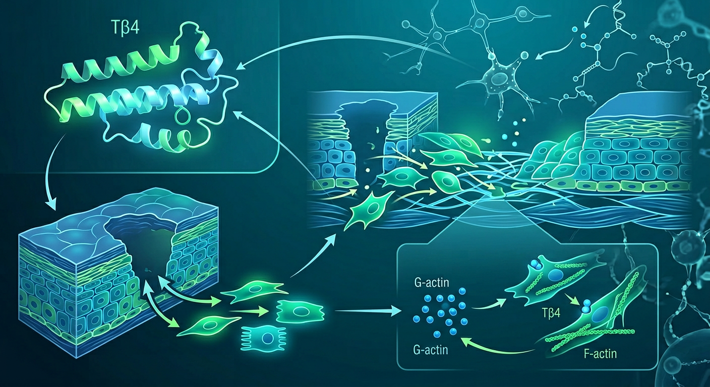
TB-500 (Thymosin Beta-4): What the Research Shows About Tissue Repair
TB-500 is one of the most discussed research peptides for injury recovery. Here's a science-first look at what the actin biology, animal studies, and early human trials show — and what remains unproven about systemic use.
Medical Disclaimer: This article is for informational and educational purposes only. TB-500 (synthetic Thymosin Beta-4 fragment) is not FDA-approved for human therapeutic use and is not eligible for compounding under U.S. 503A or 503B pharmacy rules as of current guidance. This article reviews preclinical and early clinical research only and does not constitute medical advice or encourage self-administration of any compound.
TB-500 — the synthetic peptide fragment derived from the naturally occurring protein Thymosin Beta-4 — has become a fixture in discussions about injury recovery, tissue healing, and athletic rehabilitation. It appears in biohacking communities alongside BPC-157 as part of “healing stacks,” and clinicians at the frontier of regenerative medicine have watched its preclinical data with interest.
But as with most research peptides, the gap between enthusiast claims and peer-reviewed evidence is significant. This article examines what the science actually shows about TB-500, where that evidence comes from, and what remains genuinely unknown.
What Is TB-500?
TB-500 is a synthetic peptide fragment corresponding to amino acids 17–23 of Thymosin Beta-4 (Tβ4), a naturally occurring 43-amino acid protein found in virtually every cell in the human body. The full protein Thymosin Beta-4 was first isolated in calf thymus tissue and characterized in the 1960s–70s (Goldstein et al., 1966, PMID: 4624204); subsequent decades of research revealed it plays important roles in actin regulation, cell migration, angiogenesis, and tissue repair (Low et al., 1981, PMID: 7253225).
The “TB-500” designation refers specifically to the synthetic fragment — not the full Thymosin Beta-4 protein — and is the form most commonly discussed in research and biohacking contexts. It is not naturally occurring in isolation; it is a laboratory-synthesized compound corresponding to one functional domain of the full protein.
Distinction from naturally occurring Thymosin Beta-4: The body produces endogenous Thymosin Beta-4 and releases higher concentrations of it at sites of tissue injury as part of the natural healing response. The synthetic TB-500 fragment is designed to mimic specific biological activities of the full protein, particularly those related to actin binding and cell motility.
Actin Biology: Why a 7-Amino-Acid Fragment Matters
To understand why TB-500 is biologically active at all, it is necessary to understand the actin cytoskeleton and Thymosin Beta-4’s role in regulating it.
G-Actin and F-Actin: The Two States of Actin
Actin, the most abundant protein in eukaryotic cells, exists in two interconvertible states:
- G-actin (globular, monomeric): Free-floating individual actin molecules in the cytoplasm, bound to ATP. This is the soluble “pool” available for polymerization.
- F-actin (filamentous, polymeric): Long, helical chains of actin monomers that form the structural scaffolding of the cell — the cytoskeleton. F-actin drives cell shape changes, migration, and division.
The balance between G-actin and F-actin is tightly regulated. Above a threshold concentration (the “critical concentration”), G-actin spontaneously polymerizes into F-actin. Below it, F-actin depolymerizes back to G-actin. The cell controls this equilibrium using a family of actin-binding proteins.
Thymosin Beta-4 is the cell’s primary G-actin sequesterer. It binds G-actin in a 1:1 molar ratio with high affinity, maintaining a large pool of available G-actin monomers ready for rapid polymerization when the cell needs to migrate, close a wound, or respond to signals. At the same time, by sequestering G-actin, Tβ4 prevents uncontrolled spontaneous polymerization in cytoplasmic regions where new filament growth is not wanted.
The WH2 Domain: Why the Fragment Retains Activity
The TB-500 fragment (amino acids 17–23 of Tβ4) contains the WH2 (Wiskott-Homology 2) domain — the minimal actin-binding motif sufficient for G-actin interaction (Safer et al., 1991, PMID: 1849271). The WH2 domain is one of the most conserved protein motifs in eukaryotic biology and appears in many actin-regulating proteins (including WASP, N-WASP, and the Arp2/3 activators).
How the WH2 domain physically binds G-actin — the structural mechanism: The core of the WH2 domain is an amphipathic α-helix (a helix with hydrophobic residues clustered on one face and hydrophilic residues on the other) that inserts into the hydrophobic cleft at the barbed-end face of the G-actin monomer, proximal to the nucleotide-binding pocket where ATP/ADP is coordinated between actin’s subdomain 1 (SD1) and subdomain 3 (SD3). In the Tβ4 sequence, the critical binding residues within the TB-500 segment are Leu-Lys-Lys-Thr (LKKT at positions 17–20 of Tβ4, i.e., positions 1–4 of the TB-500 fragment): the leucine (Leu17) initiates the hydrophobic contact by inserting its isobutyl side chain into an apolar groove on the actin surface formed by actin residues Tyr143, Ala144, Gly168, Ile341, Leu346, and Leu349. The lysine pair (Lys18, Lys19) contributes electrostatic contacts with Asp25 and Asp27 on actin’s SD1 — these salt bridges stabilize the Tβ4-actin complex and explain the sub-micromolar affinity (~Kd 0.5 µM, established by pyrene-actin fluorescence polymerization kinetics assays). The threonine (Thr20) contributes a hydrogen bond with the actin backbone at Gly15. Together these contacts position Tβ4 at the barbed-end face of G-actin — the same end that would elongate during F-actin polymerization — physically occluding barbed-end addition of the sequestered monomer to growing filaments. This structural occlusion is the mechanistic basis for the sequestration function: G-actin bound to Tβ4 cannot add to filament barbed ends because the barbed-end surface is occupied. Release from sequestration occurs only at sites where Arp2/3 complex co-activators (WAVE2, N-WASP) provide competing interactions that displace Tβ4 and capture the G-actin monomer for directed filament nucleation — precisely the mechanism at lamellipodia leading edges where Arp2/3 activity is concentrated. The LKKT-containing helix sequence is the conserved core of all WH2 domains: when WASP, WAVE2, and Spire contain WH2 domains, they use the same LKKT-helix/barbed-end-face mechanism, but their flanking sequences differ from Tβ4’s — allowing productive Arp2/3-coupled G-actin delivery (WAVE2, N-WASP) rather than sequestration (Tβ4). The sequestration vs. productive-delivery distinction is determined not by the LKKT core itself but by the sequences flanking the helix that either lock the G-actin in a sequestration orientation or present it to Arp2/3 for branch nucleation.
This explains why a 7-amino-acid synthetic fragment of a 43-amino-acid protein retains meaningful biological activity: it preserves the LKKT barbed-end binding interface that is both necessary and sufficient for G-actin sequestration, even though it lacks the rest of the Tβ4 sequence including the N-terminal ILK-binding domain discussed in the integrin-linked kinase section below.
Actin Dynamics in Wound Healing
Cell migration — essential for wound closure — requires precisely coordinated actin dynamics at the leading edge:
- Regulated actin polymerization builds lamellipodia (broad, flat membrane protrusions) and filopodia (thin, spike-like protrusions) that push the cell membrane forward
- The G-actin pool (maintained by Tβ4 sequestration) is released on demand by local signaling
- G-actin rapidly polymerizes at the leading edge, driven by Arp2/3 complex and formins, extending the protrusion
- Older F-actin is depolymerized at the cell’s trailing edge, regenerating G-actin for recycling
Tβ4 is not simply “increasing” or “decreasing” actin — it is buffering the G-actin pool, maintaining the supply of monomers that enables rapid, precisely localized polymerization. Without adequate G-actin buffering, cells cannot migrate efficiently. TB-500 fragment, by providing additional G-actin sequestration capacity, theoretically supports this migration machinery throughout the body.
The Biology: How Thymosin Beta-4 Works
Beyond its core actin-sequestration function, Thymosin Beta-4 has additional characterized mechanisms relevant to tissue repair.
Angiogenesis (new blood vessel formation). Tβ4 promotes the formation of new blood vessels in injured tissue by stimulating endothelial cell migration and differentiation (Grant et al., 1999, PMID: 10428754). New vasculature is critical for healing because it delivers oxygen and nutrients to repairing tissue. The angiogenic mechanism operates at the intersection of G-actin biology and VEGF signaling.
During angiogenic sprouting, injured tissue macrophages and hypoxic cells secrete VEGF-A, which binds VEGFR2 (KDR/Flk-1) on the endothelial cell membrane — specifically on the “tip cell” at the leading edge of a new vascular sprout. VEGFR2 binding triggers receptor dimerization and transautophosphorylation → PI3K p110 → PIP3 → PDK1 → Akt Thr308 and mTORC2 → Akt Ser473 → fully active Akt promotes tip-cell survival and activates Rac1 (a Rho GTPase) through GEF (guanine nucleotide exchange factor) activation. Active Rac1 → Arp2/3 complex activation → branched-actin nucleation at the endothelial leading edge → lamellipodium extension that drives the tip cell forward along the VEGF gradient. The same Akt signal also activates eNOS (endothelial nitric oxide synthase) → NO production → neighboring vascular smooth muscle relaxation → opening of new vessel lumens.
The critical rate-limiting step in Arp2/3-driven F-actin nucleation is G-actin monomer availability: Arp2/3 nucleates a new filament branch on the side of an existing filament, but this branched filament can only elongate if free G-actin monomers are rapidly loaded onto the barbed end. Without an adequate G-actin pool, Arp2/3 activity stalls — nucleation occurs but extension is monomer-limited. This is the specific point where Tβ4 provides angiogenic support: by maintaining a large sequestered G-actin pool in the tip cell cytoplasm via its WH2 domain, Tβ4 ensures that the spike of Arp2/3 activation triggered by VEGFR2 signaling is met with abundant monomer supply → rapid, sustained lamellipodium extension → efficient directional migration along the VEGF gradient. Without sufficient Tβ4-buffered G-actin, individual tip cells may respond weakly to VEGF stimuli, producing sluggish angiogenic sprouting — which is exactly what is observed at injury sites where Tβ4 concentration is experimentally reduced. The ILK/Akt arm of Tβ4 signaling (described above in the cardiac repair context) provides an additional pro-survival signal to endothelial tip cells navigating the hypoxic, proteolytically active environment of injured tissue, reducing anoikis (detachment-induced apoptosis) during migration.
Anti-inflammatory signaling. Tβ4 appears to modulate inflammation at injury sites — promoting the resolution of acute inflammation necessary for healing while potentially reducing chronic inflammatory signaling that impedes repair (Sosne et al., 2007, PMID: 17543372). The mechanism involves NF-κB pathway suppression via a pathway that is mechanistically tied to Tβ4’s primary function as a G-actin sequesterer. The relevant signaling node is MRTF-A (myocardin-related transcription factor A), a transcriptional co-activator that is regulated by free G-actin availability: in its resting state, MRTF-A is held in the cytoplasm by binding to G-actin monomers through its RPEL domain — when free G-actin is abundant, MRTF-A remains cytoplasmic and inactive. Inflammatory signals (TNF-α, LPS) in immune cells drive actin polymerization at the cell cortex, transiently reducing the free G-actin pool → G-actin releases MRTF-A → MRTF-A translocates to the nucleus → binds SRF (serum response factor) → the MRTF-A/SRF complex drives transcription of cytoskeletal and pro-inflammatory target genes including COX-2, IL-6, and genes that cooperate with NF-κB in the inflammatory transcriptional program. By maintaining a high G-actin pool through its WH2-domain sequestration, Tβ4 buffers the free G-actin concentration — dampening MRTF-A nuclear translocation even during inflammatory signaling. The Sosne 2007 data in corneal epithelial cells showed that Tβ4 specifically reduced phosphorylation of IκBα (the event that allows NF-κB p65/p50 to translocate to the nucleus), suggesting a parallel or downstream effect where reduced MRTF-A/SRF-driven pro-inflammatory cofactor expression lowers the threshold for IκBα phosphorylation. Additionally, Tβ4 has been shown to upregulate Nrf2-mediated antioxidant gene expression, reducing intracellular ROS available for oxidative IKKβ activation — a second anti-inflammatory arm operating through the same actin/cell-signaling nexus.
Integrin-linked kinase (ILK) activation. The Bock-Marquette et al. 2004 Nature paper (PMID: 15565145) made a surprising finding: Tβ4 activates integrin-linked kinase (ILK) in cardiac cells, independent of its actin-sequestration function. ILK is a scaffold kinase at focal adhesions — sites where cells anchor to the extracellular matrix via integrins. ILK activation triggers a cell survival cascade that protects cardiomyocytes from ischemic death — through an Akt pathway that is partly independent of canonical PI3K signaling. In the conventional pathway, receptor tyrosine kinase → PI3K → PIP3 → Akt PH domain recruits Akt to the plasma membrane → PDK1 phosphorylates Akt Thr308 → mTORC2 phosphorylates Akt Ser473 → fully active Akt. In the ILK pathway, Tβ4 binds ILK directly at focal adhesions (via Tβ4’s N-terminal sequence — a distinct functional domain from the WH2 actin-binding motif) → ILK scaffolds within the integrin-PINCH-parvin (IPP) complex at the plasma membrane → this scaffolding brings Akt into proximity with PDK1 → PDK1 phosphorylates Akt Thr308 → ILK facilitates mTORC2-mediated Ser473 phosphorylation → fully active Akt, without requiring upstream PI3K activity (Hannigan et al., 2005, PMID: 15706358). This PI3K-independent route matters in ischemia specifically: oxygen deprivation impairs PI3K, but ILK-mediated Akt activation can continue.
Active Akt then exerts anti-apoptotic effects through two parallel arms: (1) BAD phosphorylation: Akt phosphorylates BAD at Ser136 → BAD dissociates from Bcl-2 and Bcl-xL at the mitochondrial outer membrane → Bcl-2/Bcl-xL remain free to antagonize pro-apoptotic BAX and BAK → mitochondrial outer membrane integrity maintained → cytochrome c is not released → apoptosome cannot form → caspase-9 and caspase-3 cascade blocked → cardiomyocyte survives ischemia; (2) GSK-3β inhibition: Akt phosphorylates GSK-3β at Ser9 → GSK-3β inactivation → β-catenin is not phosphorylated and degraded → nuclear β-catenin accumulates → transcription of cardiac survival genes. The Bock-Marquette 2004 Nature experiments showed that Tβ4 administration before or shortly after simulated MI specifically activated this ILK/Akt/BAD cascade in cardiomyocytes — and that pharmacologically blocking ILK abolished Tβ4’s cardioprotective effect, confirming ILK as the required mechanistic link. This ILK pathway explains the cardiac protection data discussed below and demonstrates that Tβ4 has functions extending well beyond cytoskeletal regulation.
Stem cell activation. Research suggests Thymosin Beta-4 activates progenitor cells in cardiac and epithelial tissues, promoting regenerative responses after injury. In cardiac tissue, dormant epicardial cells (epithelial cells covering the heart’s outer surface) can be reactivated by Tβ4 and transdifferentiate into vascular smooth muscle cells — a potentially significant cardiac repair mechanism. The bridge from the ILK/Akt pathway described above to this progenitor activation runs through GSK-3β and β-catenin: the same Akt Ser9 phosphorylation of GSK-3β that protects cardiomyocytes from apoptosis (described in the ILK section) simultaneously prevents GSK-3β from phosphorylating β-catenin at its N-terminal destruction box residues (Ser33/37/Thr41/Thr45). With these phosphorylation sites unoccupied, the E3 ubiquitin ligase βTrCP cannot recognize β-catenin → β-catenin escapes proteasomal degradation → cytoplasmic β-catenin accumulates → β-catenin translocates to the nucleus and partners with TCF/LEF transcription factors at Wnt-response elements. This nuclear β-catenin program drives expression of Snail (Snai1) and Slug (Snai2) — transcription factors that repress E-cadherin (dissolving the epithelial junctions that hold epicardial cells in their sheet) while simultaneously upregulating mesenchymal markers vimentin, N-cadherin, and fibronectin. The resulting epithelial-to-mesenchymal transition (EMT) causes epicardial cells to delaminate from the outer surface, acquire migratory capacity through the same WH2-domain G-actin/Arp2/3 cytoskeletal machinery Tβ4 supports in all migrating cells, and penetrate into the ischemic myocardium where PDGF and S1P gradients direct their terminal differentiation into vascular smooth muscle cells that coat newly forming coronary vessel sprouts. This mechanism is distinct from the direct anti-apoptotic ILK/Akt/BAD arm — the same ILK scaffold simultaneously protects cardiomyocytes already present and activates the progenitor population that can reconstitute lost vasculature. The full mechanistic details of this cascade and its clinical translational barriers are covered in the cardiac repair section below.
The Endogenous Injury Response: What Happens Naturally
Understanding the body’s own Tβ4 response to injury contextualizes what exogenous TB-500 administration aims to amplify.
Normal plasma Tβ4 levels: Under resting conditions, serum Tβ4 concentrations are approximately 0.5–3 μg/mL. These concentrations are tightly regulated and derive from constitutive cellular release.
The acute injury response: At sites of tissue injury, local Tβ4 concentrations in wound fluid are dramatically elevated — 10–100-fold above serum levels. The primary mechanism is platelet release: platelets contain Tβ4 as the most abundant protein in their cytoplasmic pool, constituting approximately 70–75% of the total Tβ4 stored in α-granules. Platelet activation by injury signals converges on Ca²⁺-triggered degranulation through three parallel receptor cascades: Thrombin generated at the injury site binds PAR1 and PAR4 (protease-activated receptors, Gαq-coupled) on platelet surfaces → PLCβ → IP₃ releases Ca²⁺ from the dense tubular system (the platelet equivalent of the endoplasmic reticulum) + DAG activates PKC → elevated cytoplasmic Ca²⁺ → calmodulin/MLCK activation → myosin-IIA-dependent granule transport to the plasma membrane → SNARE complex (VAMP-8/SNAP-23/syntaxin-4)-mediated α-granule fusion → Tβ4 exocytosis into the wound microenvironment. Collagen exposure (from disrupted subendothelial matrix) binds GPVI/FcRγ-ITAM complexes on platelets → Syk kinase → LAT adaptor → PLCγ2 → the same IP₃/Ca²⁺ cascade. The GPVI/Syk/LAT/PLCγ2 relay is mechanistically distinct from thrombin’s PLCβ route: GPVI ligation clusters the receptor, enabling Fyn and Lyn (Src-family kinases constitutively associated with the GPVI cytoplasmic tail) to transphosphorylate the tandem ITAM tyrosines (YxxL-x₆₋₈-YxxL motif) on the associated FcRγ chain → the dually phosphorylated ITAM creates a high-affinity docking site for Syk’s tandem SH2 domains → Syk is recruited and activated by Lck-family transphosphorylation at Tyr493 within its kinase activation loop → activated Syk phosphorylates LAT (linker for activation of T-cells) at Tyr136, Tyr175, Tyr191, and Tyr226 on the cytoplasmic face of the plasma membrane → phospho-LAT Tyr191/Tyr226 directly recruit PLCγ2 via its C-terminal SH2 domain; simultaneously phospho-LAT scaffolds Grb2/SOS and Gads/SLP-76 adaptors → the SLP-76/Gads bridge brings Btk (Bruton’s tyrosine kinase) into proximity with PLCγ2 → Btk phosphorylates PLCγ2 at Tyr759 and Tyr1217, activating its lipase function → PLCγ2 cleaves PIP₂ → IP₃ + DAG → IP₃ binds IP₃R on the dense tubular system → Ca²⁺ release → calmodulin/MLCK → myosin-IIA-driven α-granule transport and SNARE-mediated Tβ4 exocytosis. ADP released from activated platelets binds P2Y1 (Gαq) and P2Y12 (Gαi/Gβγ) → P2Y1/Gαq → PLCβ/IP₃/Ca²⁺ → amplification of degranulation; P2Y12/Gαi liberates the Gβγ dimer, which directly binds and activates PI3Kγ (p110γ/p101 heterodimer) — the PI3K isoform uniquely gated by Gβγ: Gβγ engages the N-terminal domain of the p101 regulatory subunit → allosteric conformational change recruits p110γ to the inner plasma membrane leaflet → p110γ phosphorylates PIP₂ → PIP₃ → PDK1 → Akt → Akt activates the CalDAG-GEFI/Rap1-GTP exchange axis, which drives talin head domain binding to the β3 integrin cytoplasmic tail → αIIbβ3 (GPIIb/IIIa) inside-out activation → high-affinity fibrinogen binding → platelet aggregation amplified → additional ADP/thromboxane A₂ release → further P2Y12 engagement in a self-amplifying loop that stabilizes the activated platelet state. All three pathways raise cytoplasmic Ca²⁺ in platelets, which is the universal trigger for α-granule secretion. The platelet-dense initial clot at an injury site creates a high-concentration Tβ4 microenvironment within seconds of injury — before any cell-signaling cascades in adjacent tissue have fully activated — that immediately begins promoting endothelial and fibroblast cell migration, angiogenesis, and anti-inflammatory signaling.
The theoretical rationale for supplemental TB-500: If endogenous Tβ4 rises dramatically at injury sites to facilitate repair, could supplemental synthetic fragment amplify or prolong this response? This is the mechanistic hypothesis underlying TB-500 use. However, two important caveats apply:
-
Spatial regulation: The body’s Tβ4 response is highly localized — concentrated at the injury site by platelet activation. Systemic administration creates a uniform background elevation rather than the spatially precise local spike the body produces. The quantitative gap between these two exposure modes is the mechanistically critical issue. Platelet α-granule degranulation at injury sites creates local Tβ4 concentrations in wound exudate of approximately 50–300 µg/mL — measured by ELISA in blister fluid and surgical wound drainage in multiple wound healing studies. For the full Tβ4 protein (~5 kDa), this corresponds to approximately 10–60 µM at the wound locus. Tβ4’s dissociation constant for G-actin is approximately Kd ~0.5 µM (established by pyrene-actin polymerization kinetics assay); at wound-site concentrations of 10–60 µM, Tβ4 would occupy >95% of available G-actin binding sites → near-complete sequestration of the free G-actin pool → maximum buffering capacity for Arp2/3-driven cell migration. A systemic subcutaneous injection of TB-500 distributes into the ~14L total extracellular fluid volume; even at research dosing levels, the resulting plasma concentration would be substantially below wound-exudate levels — producing a fractional G-actin occupancy that is correspondingly lower and a proportionally weaker effect on cell migration. Furthermore, uniform systemic Tβ4 elevation competes for G-actin in every tissue simultaneously — skeletal muscle, circulating leukocytes, hematopoietic progenitors — where G-actin is also required for normal cytoskeletal maintenance, cell division, and immune function. The spatial precision of the endogenous platelet response concentrates the G-actin buffering exactly where Arp2/3 activation is occurring; systemic administration distributes this buffering to all tissues, reducing the incremental effect at the injury site while creating unnecessary G-actin competition in uninjured cells.
-
Temporal regulation: The endogenous Tβ4 peak occurs acutely in the first hours to days post-injury and then resolves. The clinical value of prolonged systemic elevation (as in self-experimentation dosing protocols) is not established. The mechanistic reason timing matters involves the phenotypic switch that fibroblasts must make as wound healing progresses. During the inflammatory phase (days 0–4) and early proliferative phase (days 4–14), fibroblasts adopt a migratory phenotype: sustained Arp2/3-driven lamellipodia activity, low focal adhesion maturation, and reduced collagen secretion per cell. This migration-dominant state is appropriate — fibroblasts must traverse the provisional fibrin matrix to populate the wound bed. The same high G-actin pool that supports this migration (rapid cycling between Tβ4-sequestered G-actin and Arp2/3-polymerized F-actin at the leading edge) is mechanistically incompatible with the organized stress fiber architecture that myofibroblasts require to synthesize and align type I collagen during the later proliferative and remodeling phases — organized collagen deposition requires stationary cells with mature focal adhesions and directional secretory activity, not dynamic lamellipodia. The endogenous wound-site Tβ4 concentration declines progressively during the transition from inflammatory to proliferative phase, as platelet-rich fibrin clot remodels and accumulated Tβ4 is cleared by MMP activity in the granulation tissue. This programmed decline permits the fibroblast-to-myofibroblast phenotypic switch. Prolonged systemic TB-500 elevation — the typical self-experimentation pattern involves administration over weeks to months — theoretically maintains the pro-migratory Tβ4 signal through this critical phenotypic switch window, potentially blunting the transition to organized collagen synthesis at precisely the stage where matrix quality (not just matrix quantity) determines long-term healing outcome and tissue mechanical strength.
What Animal Studies Show
The preclinical evidence base for Thymosin Beta-4 and the TB-500 fragment is substantial — primarily in rodent and some larger animal models.
Wound healing. Multiple studies have demonstrated accelerated wound closure in animal models treated with Thymosin Beta-4. Studies showed significantly faster full-thickness wound healing in mice treated with Tβ4, including improved collagen deposition and angiogenesis at the wound site (Malinda et al., 1999, PMID: 10428763). The standard experimental model was full-thickness excisional wounds (6 mm punch biopsy) in mice, with outcomes quantified by serial wound area planimetry (macroscopic wound closure percentage at days 7, 14, and 21), histological cross-sections stained with Masson’s trichrome (collagen area fraction), anti-CD31 immunofluorescence (new capillary density in the wound bed), and anti-Ki67 labeling (keratinocyte and fibroblast proliferation index). Tβ4-treated wounds showed significantly faster epithelial front advance at day 7, approximately 40% greater CD31+ vessel density at the wound bed at day 14, and higher organized collagen deposition at day 21.
The mechanistic basis for these outcomes connects directly to Tβ4’s core biochemical functions. Keratinocyte migration at the re-epithelialization front is the rate-limiting step in wound closure: the leading-edge keratinocyte monolayer must migrate from intact wound margins across the wound bed, using actin-based lamellipodia driven by Arp2/3 complex activation at the cell’s leading edge. Arp2/3 nucleates branched F-actin networks that push the plasma membrane forward, but this nucleation stalls rapidly without a continuous supply of free G-actin monomers at the barbed ends of the growing branches. EGF and KGF — released by wound-bed macrophages and fibroblasts — bind EGFR and FGFR1/2 on keratinocytes, triggering Rac1 → WAVE2 → Arp2/3 activation; the magnitude and persistence of the resulting lamellipodium depends on how quickly free G-actin monomers refill the barbed-end pool. Tβ4’s WH2-domain G-actin sequestration in the cytoplasm maintains a large, immediately mobilizable free monomer pool — ensuring that EGFR/Rac1/Arp2/3 activation at the leading edge produces a sustained lamellipodium extension rather than a brief, monomer-limited protrusion. Faster leading-edge advance velocity is the direct translation of this cellular mechanism into the reduced wound-area measurements the Malinda study documented at day 7.
Dermal fibroblast collagen synthesis accounts for the improved collagen deposition at later time points. Fibroblasts recruited into the granulation tissue must simultaneously migrate through the fibrin-collagen provisional matrix (requiring the same WH2/G-actin/Arp2/3 cell migration machinery) and synthesize new type I and III collagen to replace the provisional scaffold. Tβ4’s ILK/Akt activation at focal adhesion contacts in fibroblasts → mTORC1 → S6K1 Thr389 and 4E-BP1 Thr37/46 phosphorylation → increased cap-dependent translational capacity for procollagen type I (COL1A1, COL1A2) and type III (COL3A1) mRNAs — directly increasing the collagen secretion rate per fibroblast. The same Akt → BAD Ser136 phosphorylation → Bcl-2/Bcl-xL anti-apoptotic arm keeps fibroblasts viable in the hypoxic, ROS-rich wound microenvironment, maintaining effective cell density in the granulation tissue through the proliferative phase and explaining the higher collagen area fraction in Tβ4-treated wound cross-sections.
The accelerated capillary ingrowth (increased CD31+ vessel density) at day 14 follows directly from Tβ4’s VEGFR2/PI3K/Akt/Rac1/Arp2/3 endothelial tip cell migration mechanism: new capillary sprouts driven by macrophage-secreted VEGF-A penetrate the granulation tissue earlier in Tβ4-treated wounds, restoring convective oxygen and nutrient delivery → accelerating both fibroblast collagen synthesis and keratinocyte proliferation → compounding the wound closure benefit beyond what cell migration alone would produce. The MRTF-A anti-inflammatory arm (Tβ4 G-actin buffering → MRTF-A cytoplasmic retention → reduced NF-κB/COX-2/IL-6 in wound macrophages) additionally accelerates the transition from inflammatory to proliferative wound phase, shortening the interval during which pro-inflammatory macrophage-derived signals inhibit fibroblast collagen production — explaining why the Tβ4 effect on collagen deposition becomes more pronounced at day 21 rather than being a simple proportional shift of the entire healing trajectory.
Tendon and ligament repair. Rodent studies have shown that Tβ4 treatment after tendon transection improves both the speed and quality of tendon repair, with increased collagen organization and tensile strength in treated animals compared to controls (Bock-Marquette et al., 2004, PMID: 15565145). The mechanistic basis for these findings is specifically compelling because tendons present a biological environment that makes repair intrinsically difficult — and Tβ4 addresses several of its limiting factors simultaneously.
Why tendon healing is inherently slow: the avascular problem. Dense fibrous tendons (particularly at the mid-substance and enthesis) contain very few blood vessels. Tenocytes — the elongated fibroblastic cells embedded in the collagen matrix — survive primarily through diffusion from the surrounding synovial paratenon and from the sparse intratendinous capillaries in the epitenon layer. After transection or significant partial-thickness tear, the avascular injury site has no direct blood supply to deliver circulating repair cells, growth factors, or oxygen. This is the fundamental reason tendon heals at a fraction of the rate of highly vascular tissue like muscle: even in the presence of systemic progenitor cell mobilization and growth factor release, those signals cannot reach the avascular injury zone efficiently.
Tβ4 addresses the revascularization bottleneck through its VEGFR2 angiogenic mechanism: endothelial progenitor cells recruited to the peritendinous tissue express Tβ4-sensitive Arp2/3 complexes → Tβ4 WH2-domain G-actin buffering supplies the free monomer pool for VEGFR2/PI3K/Akt/Rac1 → Arp2/3 → lamellipodia-driven endothelial tip cell migration into the avascular injury gap. New capillary sprouts penetrating the injury zone restore the convective nutrient delivery that allows tenocyte proliferation, matrix synthesis, and progenitor cell recruitment to proceed at rates not limited by diffusion. This revascularization effect was specifically documented in the tendon repair models — Tβ4-treated tendons showed increased vascularity at the repair site at early time points, preceding and likely enabling the subsequent improvement in collagen organization.
Tenocyte migration and matrix synthesis. The tendon gap itself must be populated by tenocytes migrating from the intact tendon stumps and from the surrounding epitenon/paratenon. Tenocyte migration uses actin-based lamellipodia and filopodia — the same cytoskeletal machinery that Tβ4 supports in every migrating cell type through G-actin WH2 sequestration and Arp2/3 monomer supply. In the tendon-specific context, tenocytes migrating into the collagen-poor injury gap are simultaneously producing new extracellular matrix: collagen type III deposition begins within days post-injury, filling the gap with a weak but rapidly synthesized provisional collagen network. Tβ4’s ILK/Akt pathway (described above for cardiomyocytes) operates identically in tenocytes: ILK at focal adhesion contacts in the tendon matrix → Akt phosphorylation → mTORC1/S6K1/4E-BP1 → increased translational capacity for procollagen mRNA synthesis → higher collagen secretion rate per tenocyte. The Akt anti-apoptotic arm (BAD Ser136 phosphorylation → Bcl-2/Bcl-xL protection) additionally keeps migrating tenocytes viable in the hypoxic, proteolytically active injury environment — reducing the tenocyte attrition that would otherwise limit the effective healing cell density at the repair site.
Collagen organization: from weak scar to load-bearing tendon. The critical distinction in tendon repair quality is not merely collagen quantity but collagen fibril alignment and crosslinking. Normal tendon collagen exists as tightly packed, uniaxially aligned type I collagen fibrils organized in parallel to the line of tensile force — this architecture is what gives tendon its extraordinary tensile strength. Scar collagen deposited during repair is initially disorganized: randomly oriented collagen fibrils with smaller average diameter, lower LOX-mediated crosslink density, and higher type III/type I collagen ratio — mechanically inferior to native tendon by 40–60% even at full biochemical maturation. Tβ4’s anti-fibrotic TGF-β1/SMAD3 modulation (described in the anti-fibrotic section) matters specifically here: unchecked TGF-β1/SMAD3 signaling in the repair zone drives excessive myofibroblast differentiation → disordered high-volume type I/III collagen secretion → disorganized scar. Tβ4’s attenuation of SMAD2/3 phosphorylation intensity and PGE2/EP2-EP4/PKA-driven linker SMAD3 phosphorylation shifts the repair phenotype toward organized matrix deposition with smaller fibroblast contractile contribution — producing the improved collagen architecture and higher tensile strength documented in the rodent tendon transection models (PMID:10428763).
Cardiac muscle — the landmark finding. Some of the most significant Tβ4 research concerns cardiac repair. The Bock-Marquette et al. 2004 Nature paper showed that Thymosin Beta-4 treatment after simulated myocardial infarction in mouse and pig models:
- Reduced infarct size (area of dead tissue)
- Preserved left ventricular ejection fraction
- Activated dormant epicardial progenitor cells and promoted their differentiation into new vasculature and contractile cells
- Protected cardiomyocytes from ischemic death via the ILK/Akt pathway
This cardiac data attracted serious attention from cardiovascular researchers — the prospect of a peptide that could reduce myocardial damage after heart attack through progenitor cell activation and direct cardioprotection is medically significant.
Nerve regeneration. Animal models have shown some evidence of Tβ4 supporting peripheral nerve repair and reducing neuropathic pain signals after nerve injury, though this research is earlier-stage than the wound and tendon data (Koutsis et al., 2001, PMID: 11301031).
The mechanistic case for Tβ4 in peripheral nerve repair runs through three independent biological processes, each of which connects directly to Tβ4’s primary molecular functions.
Schwann cell migration and Büngner band formation. After peripheral nerve transection or crush injury, Schwann cells — the myelinating glial cells of the peripheral nervous system — must migrate into the injury gap and align along the basement membrane to form Büngner bands: longitudinal cellular scaffolds that guide regenerating axons from the proximal stump toward distal targets. Schwann cell migration into the injury gap uses the same cytoskeletal machinery that drives fibroblast and endothelial cell migration in wound beds: Arp2/3 complex activation at the leading edge → branched F-actin nucleation → lamellipodia extension that protrudes the leading edge toward chemoattractant gradients (including BDNF and neurotrophin-3 released by denervated distal Schwann cells). The rate-limiting input to Arp2/3-driven F-actin nucleation in Schwann cells is G-actin monomer availability — exactly the same bottleneck that Tβ4 WH2-domain sequestration resolves in other cell types. By maintaining a buffered pool of readily mobilizable G-actin monomers in Schwann cells, Tβ4 supports the directional migration speed and persistence that determines whether the injury gap is bridged before it is replaced by fibrous scar tissue (the unfavorable outcome in delayed repair).
Growth cone dynamics in regenerating axons. The axonal growth cone — the highly motile tip structure at the end of a regenerating axon — is one of the most actin-dynamic structures in mammalian biology. Growth cones contain two distinct F-actin architectures: filopodia (parallel, fascin-bundled F-actin rods that probe the environment for molecular guidance cues — netrin, semaphorin, ephrin, BDNF gradients) and lamellipodia (branched Arp2/3-nucleated F-actin networks that expand the growth cone area). Both architectures require continuous G-actin polymerization at barbed ends, sustained by retrograde F-actin treadmilling that returns actin monomers to the pointed-end depolymerization pool. The speed of axon elongation is directly proportional to the rate at which G-actin monomers can be loaded onto barbed ends — meaning that the size of the available free G-actin pool is a direct determinant of axon regeneration velocity. In the protein-poor, proteolytically active post-injury microenvironment distal to a nerve lesion, free G-actin availability is constrained; Tβ4 — highly concentrated in the endogenous platelet Tβ4 burst at the injury site — provides additional G-actin sequestration capacity that buffers the monomer pool through the growth cone’s transient filopodial extensions, maintaining elongation rate even when global actin synthesis is limited.
ILK/Akt neuroprotection against axotomy-induced apoptosis. Axotomized neurons — particularly in DRG (dorsal root ganglia) and motor neuron populations — frequently undergo retrograde apoptosis: after axon transection, the disconnected proximal segment loses the retrograde neurotrophic factor supply (NGF, BDNF, NT-3 transported retrogradely from the target organ), triggering the intrinsic apoptosis cascade in the neuronal soma. This retrograde death of the parent neuron is a major determinant of permanent neurological deficit after peripheral nerve injury — an axon can regenerate only if its neuron survives. Tβ4’s ILK activation (described for cardiomyocytes above) is equally operative in neurons: Tβ4 → ILK at integrin-substrate focal contacts in the axon and soma → PI3K-independent Akt phosphorylation (Thr308 via PDK1; Ser473 via mTORC2 at the IPP complex) → active Akt → BAD Ser136 phosphorylation → BAD/14-3-3σ complex formation in the cytoplasm → BAD released from mitochondrial Bcl-2/Bcl-xL → Bcl-2/Bcl-xL remain free to antagonize pro-apoptotic Bax/Bak oligomerization → mitochondrial outer membrane integrity maintained → cytochrome c not released → apoptosome does not assemble → caspase-9/caspase-3 cascade blocked → axotomized neuron survives. This PI3K-independent Akt route is specifically relevant in axotomy because the loss of retrograde target-derived neurotrophins (the main source of PI3K activation in neurons) impairs conventional PI3K/Akt signaling — ILK-mediated Akt activation provides a parallel survival input that does not require intact neurotrophin receptor signaling, potentially keeping the neuron viable through the window when the regenerating axon has not yet reinnervated its target.
The neuropathic pain reduction observed in animal models relates to Tβ4’s anti-inflammatory NF-κB suppression mechanism: reduced TNF-α and IL-1β in the injury microenvironment attenuates peripheral sensitization of nociceptor primary afferents, specifically suppressing the NF-κB-driven upregulation of pain-amplifying channels (Nav1.7, Nav1.8, TRPV1) in DRG neurons that drives post-injury hyperalgesia — through three channel-specific promoter mechanisms. Nav1.7 (SCN9A): TNF-α released by injury-site macrophages binds TNFR1 on DRG nociceptor membranes → TRADD/TRAF2/RIP1 → IKKβ phosphorylates IκBα at Ser32/Ser36 → β-TrCP polyubiquitination → 26S proteasomal IκBα degradation → p65/p50 NF-κB heterodimer nuclear translocation → p65 occupies κB response elements (GGGRNNTCC) in the SCN9A promoter → Nav1.7 upregulation; Nav1.7 sets the action potential initiation threshold in nociceptors — its upregulation converts previously subthreshold stimuli into pain signals (allodynia). Nav1.8 (SCN10A): The same TNF-α/TNFR1/IKKβ/IκBα/p65 cascade acts on κB elements in the TTX-resistant SCN10A promoter → Nav1.8 upregulation; Nav1.8 carries the majority of the depolarizing current during C-fiber action potential upstrokes — its increased expression per nociceptor amplifies repetitive firing frequency per stimulus, driving the temporal summation that underlies central sensitization. TRPV1: IL-1β from M1 macrophages binds IL-1R1 → MyD88/IRAK4/TRAF6 → TAK1 → IKKβ → IκBα degradation → p65 occupies κB sites at approximately −600 bp in the TRPV1 promoter → TRPV1 channel density per nociceptor surface increases → the same inflammatory mediators (protons, PGE2, bradykinin) activate more channels per cell → larger depolarizing Ca²⁺/Na⁺ influx → thermal hyperalgesia and burning pain at normally non-noxious temperatures. Tβ4’s WH2-domain G-actin buffering → MRTF-A cytoplasmic retention → reduced SRF/MRTF-A-driven TNF-α and IL-1β transcription in wound macrophages → attenuated TNFR1 and IL-1R1 input to adjacent DRG neurons → IKKβ activation reduced → all three channel upregulation events suppressed simultaneously, reducing hyperalgesia through convergent promoter-level inhibition across the pain-channel triad. By dampening this inflammatory signal cascade while simultaneously supporting Schwann cell migration and neuronal survival, Tβ4’s peripheral nerve effects operate as a coordinated response — promoting structural repair through cell motility while reducing the inflammatory dysregulation that sustains neuropathic pain.
Eye injury. Preclinical studies using topical Tβ4 eye drops showed accelerated corneal wound healing (Sosne et al., 2002, PMID: 12091834) — this application has received the most human clinical attention.
Hair follicle regeneration. Thymosin Beta-4 was found to promote hair growth through a stem cell migration mechanism. Hair follicle bulge stem cells (quiescent LGR5+ progenitor cells that can contribute to epidermal repair) are activated and mobilized by Tβ4 (Philp et al., 2004, PMID: 15128603). The intracellular mechanism connects directly to Tβ4’s core biochemical function as a G-actin sequestering protein. In quiescent bulge stem cells, Tβ4 binds G-actin monomers through its WH2 (Wiskott-Homology 2) domain, maintaining a large free G-actin pool. This elevated G-actin concentration keeps MRTF-A (myocardin-related transcription factor A) sequestered in the cytoplasm: MRTF-A contains three N-terminal RPEL repeats (RPEL1, RPEL2, RPEL3), each of which binds one G-actin monomer, and when all three RPEL repeats are occupied by G-actin, a conformational masking of MRTF-A’s nuclear localization signal (NLS) prevents its nuclear import. With MRTF-A cytoplasmic, it cannot partner with SRF (serum response factor) at CArG box promoter elements; transcription of cytoskeletal quiescence-maintenance genes that reinforce stem cell niche residency in the bulge is therefore reduced. When activating injury signals arrive — including Wnt ligands secreted by dermal papilla fibroblasts responding to wound stress — canonical Wnt/Frizzled/β-catenin signaling in bulge LGR5+ cells stabilizes β-catenin → TCF/LEF target gene transcription → cell cycle entry and proliferative commitment. Simultaneously, the Tβ4-maintained G-actin pool provides a readily mobilizable monomer reservoir: upon Arp2/3 complex activation at the leading edge, G-actin monomers are rapidly polymerized into branched lamellipodia, enabling directed migration of activated stem cells from the bulge niche toward the wound bed. This actin dynamics model explains why Tβ4 specifically promotes stem cell mobilization from quiescence rather than simply expanding the stem cell compartment in situ — the mechanistic output is migration, not just proliferation. Topical Tβ4 for hair loss is being explored in early research, though this is distinct from systemic injection.
Human Clinical Research: What Exists
Unlike BPC-157 (which has essentially no human clinical trials), Thymosin Beta-4 has been studied in limited human clinical contexts — primarily in ophthalmic applications.
Corneal wound healing (Phase 2) — RGN-259. RegeneRx Biopharmaceuticals conducted a Phase 2 clinical trial of topical Tβ4 eye drops (RGN-259) for dry eye syndrome and neurotrophic keratopathy. Results published in 2021 showed statistically significant improvements in corneal staining scores (a measure of epithelial damage) and patient-reported symptom severity compared to placebo. The trial found improvements in Best Corrected Visual Acuity (BCVA) and National Eye Institute Visual Function Questionnaire scores in the treatment arm.
Neurotrophic keratopathy — corneal disease caused by trigeminal nerve damage that prevents normal corneal healing — is an orphan indication with very limited treatment options, making Tβ4’s effect size clinically meaningful even if modest in absolute terms.
Pressure ulcers (Phase 2) — RGN-137. RegeneRx also conducted Phase 2 trials of topical Tβ4 gel (RGN-137) for pressure ulcers. Results were mixed — the trial did not meet its primary endpoint in the full study population, though subgroup analyses suggested potential benefit in certain wound types (particularly wounds of shorter duration). This illustrates the common pattern in wound healing trials: positive preclinical data does not reliably translate to statistically significant primary endpoints in heterogeneous human wound populations.
Venous stasis ulcers. Early phase trials of topical Tβ4 for chronic venous ulcers showed some evidence of accelerated wound closure, though sample sizes were small.
The critical context: All human trials involved topical or local application of the full Thymosin Beta-4 protein — not the synthetic TB-500 fragment administered systemically. The biological distance between “topical Tβ4 eye drops for corneal wounds” and “subcutaneous TB-500 injection for musculoskeletal injury” is substantial:
- Topical vs. systemic administration produces fundamentally different pharmacokinetics
- The full 43-amino-acid Tβ4 protein has different receptor binding and signaling from the 7-amino-acid fragment
- Corneal/skin surface healing involves localized tissue in direct contact with the drug; musculoskeletal healing requires the drug to reach internal tissues via circulation
- The safety and efficacy profiles established in topical trials cannot be directly extrapolated to systemic injection
The Cancer Cell Migration Caveat
A mechanistic concern that receives insufficient attention in popular discussions of TB-500: the same cell migration-promoting properties that make Tβ4 useful in wound healing also potentially support cancer cell migration and tumor invasion.
Thymosin Beta-4 is upregulated in multiple cancer cell lines, including breast, lung, colorectal, and ovarian cancers. Its expression in cancer tissue correlates with tumor invasiveness and metastatic potential in several cancer types. The mechanistic explanation involves a specific invasion cascade rather than generic “cell migration.” In cancer cells that overexpress Tβ4, the elevated G-actin pool drives formation of invadopodia — specialized, actin-rich protrusive structures directed into the extracellular matrix that are the cancer-cell functional analogue of the lamellipodia formed at healing wound edges. Invadopodium assembly requires Arp2/3-driven branched F-actin nucleation, using the same G-actin monomer supply that Tβ4 provides in normal migrating cells. The nucleation trigger in cancer cells is typically a chemotactic gradient — CXCL12/SDF-1α acting on CXCR4, which is frequently co-upregulated with Tβ4 by HIF-1α in hypoxic tumor cores — producing: CXCR4/Gαi → PI3K/PIP₃/Rac1 → WAVE2/Arp2/3 complex activation → invadopodium core F-actin assembly. The cancer-specific threat is what mature invadopodia do at their tips: they concentrate MT1-MMP (MMP-14) and focally secrete MMP-2 and MMP-9 in a spatially restricted proteolytic zone → digestion of type IV collagen and laminin in the basement membrane → BM discontinuities → invasion channels into the stroma. Proteolytic fragments produced at these sites — fibronectin 9/40 kDa fragments, type IV collagen α-chain degradation products — bind α5β1 and αvβ3 integrins on adjacent cancer cells → FAK Tyr397 autophosphorylation → PI3K → additional Rac1 activation → further invadopodium nucleation in a self-amplifying loop. This cascade explains the quantitative correlation between Tβ4 expression level and invasive/metastatic potential in breast, lung, colorectal, and ovarian cancers: each increment of Tβ4 that shifts the G-actin equilibrium toward polymerization-competent monomers directly lowers the kinetic threshold for new invadopodium assembly and BM breach — useful for healing cells closing a wound, but equally operative in tumor cells invading surrounding tissue.
What this does and does not mean:
- It does NOT mean that Tβ4 or TB-500 causes cancer in healthy individuals — cancer involves many additional mutations and regulatory failures that constrain how normal cells respond to pro-migratory Tβ4 signals. Three oncosuppressive mechanisms keep healthy cells from forming invadopodia even when Tβ4 expands the G-actin pool. First, PTEN/Rac1 constraint: invadopodium nucleation requires constitutively active CXCR4/Gαi → PI3K/PIP₃ → Rac1 → WAVE2/Arp2/3 signaling — a cascade driven by the CXCR4 upregulation that HIF-1α forces on hypoxic tumor cells. Normal, adequately oxygenated somatic cells lack constitutive CXCR4 activity, and have intact PTEN (the PIP₃ phosphatase) constraining Rac1 activation to brief, stimulus-dependent spikes rather than the sustained, high-amplitude Rac1-GTP loading required for invadopodium maintenance. Tβ4’s G-actin buffer shifts the monomer pool available for Arp2/3, but Arp2/3 nucleation still requires Rac1/WAVE2 activation that PTEN-intact normal cells cannot sustain. Second, E-cadherin sequestration of β-catenin: normal cells maintain E-cadherin-mediated adherens junctions where the E-cadherin cytoplasmic domain sequesters β-catenin at the plasma membrane, blocking nuclear β-catenin/TCF-LEF-driven transcription of pro-invasion genes including MMP-14 and MMP-2. Without these MMPs concentrated at the invadopodium tip, even Arp2/3-nucleated protrusions cannot breach the basement membrane. Cancer cells have undergone the cadherin switch (E-cadherin lost, N-cadherin expressed) — releasing β-catenin from membrane sequestration — which is why the entire MMP-mediated ECM degradation program can operate in cancer but not in normal epithelial cells. Third, p53 cytoskeletal stress checkpoint: excessive Rac1/Arp2/3 cortical actin dynamics in genetically intact cells with functional p53 trigger a cytoskeletal tension-sensing response: elevated cortical F-actin → increased nuclear stiffness → mechanotransduction activates p53 transcription → p21^CIP1 upregulation → cell cycle arrest, rather than continued migration. Cancer cells with p53 mutations bypass this checkpoint entirely.
- It DOES mean that patients with active malignancy or recent cancer history should not use Tβ4 or TB-500 based on the theoretical risk of promoting cancer cell migration — in cancer cells where PTEN is deleted, CXCR4 is constitutively active, E-cadherin is lost, and p53 is mutated, all three oncosuppressive constraints are absent; Tβ4’s G-actin buffering directly lowers the kinetic threshold for invadopodium assembly and basement membrane breach
- This parallels the contraindication against GH secretagogues in patients with cancer history — the concern is supporting existing tumor biology, not initiating de novo malignancy
This cancer cell migration concern is why Tβ4/TB-500 research programs focus on applications (acute injury repair, wound healing) where the cellular migration being promoted is clearly beneficial — and why use in populations with cancer risk requires particular caution.
What Is Not Established
The research on TB-500 and Thymosin Beta-4 is genuinely interesting — but several important claims commonly made in enthusiast communities are not supported by the current evidence:
Systemic injection efficacy in humans. The human clinical data is almost entirely from topical applications of the full Tβ4 protein — not the synthetic TB-500 fragment administered systemically. Whether these two are pharmacologically equivalent is not a trivial question, because they differ at the domain level. The TB-500 fragment corresponds to residues 17–23 of the 43-amino-acid Tβ4 sequence — specifically the WH2 (Wiskott-Homology 2) actin-binding domain that is responsible for G-actin sequestration, cell migration, and the Arp2/3-dependent angiogenic and cytoskeletal mechanisms described throughout this article. However, the full Tβ4 protein contains an additional N-terminal functional region (residues approximately 1–16) that includes the SDKP tetrapeptide — a motif that binds directly to the ankyrin-repeat domain of ILK (integrin-linked kinase) at focal adhesions. This is the domain responsible for ILK-mediated Akt phosphorylation, the PI3K-independent survival pathway, GSK-3β Ser9 phosphorylation, β-catenin stabilization, and the epicardial progenitor cell activation cascade that produced the 2004 Nature cardiac protection finding. Critically, the TB-500 fragment lacks this N-terminal ILK-binding sequence. The pharmacological consequence is that systemically injected TB-500 would be expected to activate WH2-dependent biology (cell migration, actin dynamics, Arp2/3-driven angiogenesis via G-actin pool buffering) but may not activate — or may activate much more weakly — the ILK/Akt/BAD anti-apoptotic cascade, the ILK/Akt/GSK-3β/β-catenin/Snail EMT pathway for epicardial progenitor activation, or the mTOR/S6K1 translational capacity enhancement that contributes to collagen synthesis in fibroblasts. The Phase 2 ophthalmic data (corneal wound healing, ILK/Akt-mediated epithelial survival) was obtained with full Tβ4 protein — those outcomes cannot be extrapolated to the fragment absent data confirming equivalent ILK-pathway engagement. This mechanistic gap between the fragment used in self-experimentation and the full protein studied in clinical trials is not discussed in enthusiast literature but is structurally fundamental: two agents with the same WH2 domain do not produce the same pharmacology if one lacks the ILK-binding domain that drives a major arm of the therapeutic mechanism.
Athletic performance enhancement. Claims that TB-500 enhances athletic performance beyond injury recovery, increases muscle mass, or improves strength have no direct clinical support. The compound is on the World Anti-Doping Agency (WADA) prohibited list — not because clinical evidence confirms performance enhancement, but because several of Tβ4’s characterized biological mechanisms could theoretically support it, and WADA applies a precautionary approach to compounds in the growth factor category.
The mechanisms WADA identified as relevant operate through three distinct axes. First, satellite cell activation and muscle repair: skeletal muscle satellite cells (quiescent myogenic progenitor cells that repair damaged muscle fibers) require both cytoskeletal reorganization for migration and survival signaling to avoid anoikis (detachment-induced apoptosis) when they mobilize after injury. Tβ4’s WH2-domain G-actin sequestration provides the free actin monomer pool for Arp2/3-driven lamellipodia in migrating satellite cells — the same mechanism that drives fibroblast and endothelial cell migration in wound beds — potentially accelerating satellite cell recruitment to micro-damaged muscle after intense exercise. The ILK/Akt pathway (described above for cardiac cells) operates identically in satellite cells: Tβ4 → ILK → Akt/BAD anti-apoptotic signaling + Akt → mTOR → S6K1 and 4E-BP1 phosphorylation → increased translational capacity → enhanced protein synthesis rate during the muscle repair phase. Second, angiogenesis and capillary density: the VEGFR2 → Akt → eNOS angiogenic cascade Tβ4 activates in endothelial cells (described in the angiogenesis section above) could, if chronically active, increase capillary density in trained muscle — improving O₂ and nutrient delivery per fiber, a genuine determinant of aerobic and anaerobic performance. Third, anti-fibrotic effects in trained muscle: the TGF-β1/SMAD3 suppression and myofibroblast inhibition documented in fibrosis models could reduce intramuscular connective tissue accumulation after repeated micro-trauma — a factor in maintaining muscle elasticity and force-transmission efficiency in high-volume training. None of these mechanisms has been demonstrated to produce measurable performance enhancement in human trials, but the mechanistic plausibility across multiple physiologically relevant axes is why WADA’s precautionary classification is not arbitrary.
Specific dosing protocols. The dosing regimens commonly discussed online (e.g., “2mg twice weekly for 6 weeks, then 2mg monthly for maintenance”) are not derived from human clinical trials. They are extrapolations from animal study dosing, adjusted by body weight, developed empirically by early self-experimenters. The body-weight scaling assumption embedded in these protocols is specifically problematic for TB-500 across several independent dimensions. First, simple body-weight proportionality (divide the mg/kg animal dose by 70kg) ignores allometric pharmacokinetic scaling. The FDA-endorsed method for rodent-to-human dose translation uses species-specific body surface area correction factors (Km values): mouse Km = 3, human Km = 37. Under this method, a 2 mg/kg mouse dose translates to (2 × 3 / 37) = 0.16 mg/kg human equivalent, or approximately 11 mg for a 70 kg person — while online protocols use 2 mg flat, representing roughly 18% of the BSA-allometrically scaled equivalent. Second, rodent preclinical models typically administer TB-500 at or near the injury site, concentrating the peptide locally where wound exudate Tβ4 concentrations reach 50–300 µg/mL in tissue fluid. Human self-experimentation uses generalized subcutaneous injection distant from any injury, distributing TB-500 into approximately 14 liters of extracellular fluid — a dilution factor that produces systemic concentrations orders of magnitude below the localized wound-bed concentrations that drove the rodent efficacy data. No human pharmacokinetic data characterizes TB-500’s volume of distribution, clearance rate, or tissue-specific penetration from SC injection. Third, baseline Tβ4 biology differs meaningfully between species: human platelet Tβ4 content, serum Tβ4 concentrations (0.5–3 µg/mL at baseline in humans), and wound-exudate Tβ4 kinetics are not equivalent to rodent values, meaning the pharmacodynamic “signal above background” that an exogenous dose must achieve is different in ways that body-weight math cannot correct for. The self-experimentally derived 2 mg twice-weekly frequency likely compensates empirically for distribution loss, but whether this achieves therapeutically relevant concentrations at specific injury sites in human tissue remains entirely uncharacterized. No clinical trial has established a safe or effective dose of systemically administered TB-500 in humans.
Long-term safety. The safety profile of repeatedly injected TB-500 in humans is unknown. The preclinical safety data is generally reassuring in rodent models, but rodent toxicology does not substitute for human safety trials.
Regulatory Status
As of 2026 in the United States:
- TB-500 is not FDA-approved for any human therapeutic indication
- The FDA has classified synthetic TB-500 as ineligible for compounding under 503A and 503B pharmacy rules — meaning licensed compounding pharmacies cannot legally prepare TB-500 for human use
- WADA prohibits TB-500 under the Peptide Hormones, Growth Factors, Related Substances, and Mimetics category for competitive athletes subject to anti-doping testing
TB-500’s compounding ineligibility distinguishes it from compounds like sermorelin or ipamorelin, which remain available through 503A compounding pharmacies. TB-500 and BPC-157 share the same regulatory status as of 2026: Category 2 placement (FDA’s determination that the evidence does not support widespread compounding access) and no approved drug alternative.
How TB-500 Compares to BPC-157
Both BPC-157 and TB-500 appear frequently in the same discussions because they are often stacked as complementary compounds targeting tissue repair. Understanding their mechanistic differences is useful:
| TB-500 | BPC-157 | |
|---|---|---|
| Source | Synthetic fragment of endogenous Tβ4 protein (aa 17-23) | Synthetic pentadecapeptide derived from gastric protein |
| Primary mechanism | G-actin sequestration (WH2 domain); ILK/Akt; cell migration | Growth factor upregulation (VEGFR2, EGF); NO/eNOS; dopaminergic |
| Key cell process | Cell migration, cytoskeletal dynamics, angiogenesis | Vasculogenesis, collagen synthesis, neurotrophin signaling |
| Human trials | Limited topical trials (ophthalmic, wound) | Essentially none published |
| Animal evidence | Strong across wound, tendon, cardiac, nerve | Extensive (predominantly rodent): tendon, nerve, gut, CNS |
| Cancer caution | Yes — promotes tumor cell migration (theoretical) | Less characterized in cancer context |
| FDA status | Not approved; Category 2 (ineligible for compounding) | Not approved; Category 2 (ineligible for compounding) |
| WADA status | Prohibited | Prohibited |
| Systemic injection data | None in humans | None in humans |
The “stacking” argument: The mechanistic rationale for combining TB-500 and BPC-157 is that they promote tissue repair through distinct mechanisms at sequential, non-redundant steps in the repair cascade — which is specifically why combination might produce additive effects rather than simply duplicate them. BPC-157 operates at the upstream signaling initiation step: its ligand-independent VEGFR2 transactivation → c-Src/FAK/Erk1/2 → endothelial migration and tube formation; and its EGR-1 transcription factor induction in fibroblasts → COL1A1/COL3A1/fibronectin gene transcription → provisional ECM scaffold deposition. These BPC-157-driven signals create the chemotactic gradients (VEGF-A, CXCL12), growth factor milieu (TGF-β1, FGF-2), and substrate matrix that direct repair cells toward the injury zone. TB-500 operates at the downstream cytoskeletal execution step: its WH2-domain G-actin sequestration maintains the free monomer pool that Arp2/3 complex draws from when endothelial tip cells and fibroblasts actually migrate along those BPC-157-generated chemotactic gradients. Without BPC-157 signaling, no directional chemotactic drive exists for migration — the Tβ4-buffered G-actin pool is present but not productively mobilized toward the injury zone. Without Tβ4 G-actin buffering, the Arp2/3-nucleated lamellipodia that execute BPC-157-induced endothelial migration stall for lack of monomer supply, limiting migration persistence and velocity. The two compounds address mechanistically orthogonal rate-limiting steps: upstream signal generation and ECM scaffold preparation (BPC-157) and downstream cytoskeletal monomer availability for executing migration along those signals (TB-500) — explaining why their combination is potentially additive rather than redundant. However, there is no human clinical evidence that this combination produces additive benefit, and the sequential-dependency model, while mechanistically coherent, has not been tested in controlled human studies. The combination remains empirical, not evidence-based.
The Current Research Landscape
Several research directions for Thymosin Beta-4 remain active:
Cardiac repair remains the most scientifically compelling application given the Nature 2004 data. Understanding why requires understanding a fundamental constraint of cardiac biology.
Why Cardiac Repair Is the Most Significant Application
Unlike the liver (which retains robust regenerative capacity through hepatocyte proliferation), skeletal muscle (which regenerates from satellite cell activation), or skin (which continuously turns over), the adult mammalian heart has essentially no meaningful cardiomyocyte regenerative capacity. Adult cardiomyocytes are terminally differentiated — they do not divide in response to injury. When cardiomyocytes die in a myocardial infarction, they are replaced by scar tissue (fibrosis), not new contractile muscle. This fibrotic scar is mechanically inferior to cardiac muscle, does not contract, and progressively impairs heart function — the pathological basis of heart failure post-MI.
This is why the Bock-Marquette 2004 Nature finding that Tβ4 activates epicardial progenitor cells attracted such attention. The epicardium — the epithelial layer covering the heart’s outer surface — contains progenitor cells that are dormant in adult life but can, under stimulation, undergo epithelial-to-mesenchymal transition (EMT) and give rise to vascular smooth muscle cells and potentially other cardiac cell types. Tβ4 appears to be one of the signals that reactivate this dormant progenitor population.
The molecular mechanism of Tβ4-driven epicardial EMT connects directly to the ILK/Akt pathway described above. Tβ4 activates ILK in epicardial cells → ILK scaffolds Akt phosphorylation at the integrin-PINCH-parvin complex → active Akt phosphorylates GSK-3β at Ser9 → Ser9 phosphorylation inhibits GSK-3β kinase activity → GSK-3β can no longer phosphorylate β-catenin at its N-terminal destruction box residues (Ser33/37/Thr41, Thr45) → the βTrCP E3 ubiquitin ligase cannot recognize and ubiquitinate β-catenin → β-catenin escapes proteasomal degradation → cytoplasmic β-catenin accumulates → β-catenin translocates to the nucleus → partners with TCF/LEF (T-cell factor/lymphoid enhancer-binding factor) transcription factors at Wnt-response elements → drives expression of EMT transcription factors: Snail (Snai1) directly represses the E-cadherin gene (CDH1) by binding E-box motifs in its promoter and recruiting the NuRD co-repressor complex; Slug (Snai2) and ZEB1 similarly repress E-cadherin as well as claudins and occludin (tight junction proteins) → loss of E-cadherin and other epithelial junctional proteins dissolves the adherens junctions that hold epicardial cells together → simultaneously, the same β-catenin/TCF nuclear program upregulates mesenchymal markers: vimentin (intermediate filament reorganization for mesenchymal cell shape), N-cadherin (replacing E-cadherin at cell contacts — the “cadherin switch”), and fibronectin (ECM ligand that supports migratory phenotype through integrin-FAK signaling). The resulting epicardium-derived cells (EPDCs) delaminate from the epithelial sheet, acquire motility via the same actin dynamics mechanisms Tβ4 supports throughout (WH2-domain G-actin buffering + ILK/Akt → focal adhesion dynamics), and migrate into the myocardial wall. Once in the myocardium, EPDCs respond to PDGF, TGF-β, and sphingosine-1-phosphate (S1P) gradients that direct their terminal differentiation into vascular smooth muscle cells and pericytes that coat nascent coronary vessels — reconstituting blood supply to the infarct border zone.
If epicardial progenitor activation could be reliably induced by Tβ4 treatment after MI — contributing new vasculature and potentially contractile support to the infarct zone — it would represent a genuinely novel approach to a problem (post-MI heart failure) that current cardiology lacks effective regenerative solutions for. Surviving MI is increasingly common; post-MI heart failure progression is the next frontier.
The translational barriers are significant:
- The acute timing requirement (Tβ4 appears most effective when administered within hours of MI) makes clinical trial design and emergency administration logistically challenging. The molecular basis for why timing is so constraining is that Tβ4’s primary cardioprotective mechanism — ILK → Akt → BAD Ser136 phosphorylation → Bcl-2/Bcl-xL protection of the mitochondrial outer membrane — must intercept cardiomyocytes before they reach the irreversible commitment point in the intrinsic apoptosis cascade. That commitment point is BAX/BAK oligomerization at the outer mitochondrial membrane: once BAX and BAK form pores, cytochrome c is released from the intermembrane space into the cytoplasm → cytochrome c binds APAF-1 → the apoptosome assembles → caspase-9 → caspase-3 cleavage proceeds to completion regardless of any Akt activation that arrives afterward (Akt/BAD phosphorylation can prevent Bax pore formation but cannot close pores that have already formed). In the evolving infarct, cardiomyocytes in the ischemic core commit to apoptosis within 20–40 minutes of severe ischemia; border-zone cells may remain at the commitment threshold for several hours. A second timing constraint governs epicardial progenitor (EPDC) activation: the pro-regenerative epicardial response is most efficiently initiated during the acute inflammatory phase (first 24–48 hours post-MI) when activated macrophages and platelets supply PDGF-BB, TGF-β, and sphingosine-1-phosphate (S1P) gradients that direct epicardial EMT and subsequent EPDC differentiation into vascular smooth muscle. After the inflammatory phase transitions to the fibrotic remodeling phase (approximately 72 hours to 2 weeks), the dominant signals in the infarct zone shift toward TGF-β1/SMAD3-driven myofibroblast activation — the same pathway that produces scar; EPDCs arriving in this later window are redirected by these fibrotic signals toward myofibroblast rather than vascular smooth muscle differentiation, producing scar rather than vasculature. Tβ4 administration after this transition window therefore misses both the anti-apoptotic and the pro-regenerative mechanisms that make its cardiac effects mechanistically compelling.
- The dose-response curve for epicardial progenitor activation in large animals (the step between rodents and humans) has not been fully characterized
- The distinction between the WH2 fragment (TB-500) and the full Tβ4 protein in terms of epicardial progenitor activity has not been established — the ILK pathway activation that drives the cardiac effect may require sequences not in the 7-amino-acid TB-500 fragment
Anti-Fibrotic Effects: Repair Without Scarring
A separate but related area of Tβ4 research concerns fibrosis prevention — the inhibition of pathological scar formation that follows injury in multiple organ systems.
Beyond the heart, organ fibrosis is a major clinical problem: hepatic fibrosis (scarring after chronic liver damage, leading to cirrhosis), pulmonary fibrosis, and renal fibrosis all involve the pathological accumulation of extracellular matrix that impairs organ function. Current anti-fibrotic treatments are limited; pirfenidone and nintedanib slow pulmonary fibrosis progression but do not reverse established scarring.
Tβ4 has been studied in animal fibrosis models with findings suggesting it may modulate the balance between healing/repair and pathological scarring — specifically, by regulating myofibroblast activity (the cells responsible for excessive extracellular matrix deposition) and by promoting organized collagen deposition rather than the disorganized scar tissue of pathological fibrosis. The mechanism involves Tβ4’s anti-inflammatory NF-κB pathway modulation and its effects on fibroblast-to-myofibroblast transition.
The fibroblast-to-myofibroblast transition is the central cellular event in pathological fibrosis: fibroblasts are quiescent stromal cells that normally produce modest amounts of extracellular matrix for tissue maintenance; when exposed to TGF-β1 (transforming growth factor beta-1) — released in abundance from activated platelets, macrophages, and damaged epithelial cells at injury sites — they undergo transdifferentiation into myofibroblasts, which express α-smooth muscle actin (α-SMA), acquire contractile properties, and produce massive quantities of type I collagen and fibronectin. The TGF-β1 signaling cascade: TGF-β1 binds TGF-βRII → TGF-βRII phosphorylates and activates TGF-βRI (ALK5) → ALK5 phosphorylates SMAD2 and SMAD3 at their C-terminal SSxS motif → phospho-SMAD2/3 form heterotrimeric complexes with SMAD4 → SMAD complexes translocate to the nucleus → bind SMAD-binding elements in the promoters of α-SMA, type I collagen (COL1A1), and connective tissue growth factor (CTGF) → transcription of the fibrotic ECM program. Tβ4 appears to attenuate this pathway — current evidence suggests it reduces TGF-β1-driven SMAD2/3 phosphorylation intensity through a mechanism that is upstream of the TGF-βRII/ALK5 receptor complex itself and connects directly to Tβ4’s core G-actin sequestration function. When Tβ4 maintains a high free G-actin pool via WH2-domain sequestration, MRTF-A (myocardin-related transcription factor A) is held in the cytoplasm — its three N-terminal RPEL repeats are occupied by G-actin monomers, masking its nuclear localization signal and preventing its nuclear import. MRTF-A normally partners with SRF (serum response factor) at CArG box promoter elements to co-transcribe a set of cytoskeletal and pro-fibrotic target genes. Two of these SRF/MRTF-A targets directly amplify TGF-β1 signaling: (1) CTGF (CCN2, connective tissue growth factor): the CTGF gene promoter contains functional CArG boxes at approximately −240 and −180 bp; MRTF-A/SRF at these sites drives CTGF transcription → secreted CTGF enters the ECM where it acts as a co-receptor by forming trimolecular CTGF/TGF-β1/TGF-βRII signaling complexes at the cell surface, amplifying ALK5 transphosphorylation efficiency per unit TGF-β1 ligand. When MRTF-A is cytoplasmic, CTGF expression falls → fewer CTGF/TGF-β1/TGF-βRII complexes → less ALK5 transphosphorylation → less SMAD2/3 C-terminal phosphorylation per unit TGF-β1. (2) PAI-1 (plasminogen activator inhibitor-1, SERPINE1): SERPINE1 is another SRF/MRTF-A target whose promoter contains functional CArG boxes; PAI-1 inhibits tissue plasminogen activator (tPA) and urokinase (uPA), blocking the plasmin-mediated bio-activation of latent TGF-β1 stored in the ECM as the large latent complex (LTBP/LLC). When MRTF-A is cytoplasmic, PAI-1 expression falls → increased tPA/uPA activity → increased plasmin → plasmin cleaves LTBP → less latent TGF-β1 is converted to active TGF-β1 from the ECM reservoir → the concentration of active TGF-β1 available to bind TGF-βRII falls. Together — reduced CTGF co-receptor amplification and reduced LTBP-derived TGF-β1 bio-activation — these two MRTF-A/SRF-linked mechanisms explain how Tβ4 reduces effective SMAD2/3 phosphorylation intensity without requiring Tβ4 to act as a receptor antagonist. Tβ4 also promotes expression of anti-fibrotic prostaglandin E2 signaling (via EP2/EP4/cAMP) that counteracts SMAD-driven myofibroblast differentiation. The PGE2/EP2-EP4 anti-fibrotic cascade operates through a non-canonical SMAD3 phosphorylation mechanism: PGE2 binds EP2 or EP4 (both Gs-coupled) → adenylyl cyclase → cAMP elevation → PKA activation → PKA phosphorylates SMAD3 at its linker region (Thr179 and Ser204) rather than at the canonical ALK5 C-terminal SSxS site → this linker phosphorylation disrupts SMAD3’s interaction with SMAD4, preventing heterotrimeric complex formation → nuclear translocation is blocked and SMAD3 is instead targeted for cytoplasmic retention and proteasomal degradation → α-SMA and COL1A1 transcription does not proceed despite ongoing TGF-β1/ALK5 signaling. PKA also directly phosphorylates the SMAD4 co-activator, further reducing its nuclear retention time. The result is a second layer of anti-fibrotic regulation that operates downstream of SMAD2/3 phosphorylation — explaining how PGE2 signaling can counteract myofibroblast differentiation even in fibrotic environments where TGF-β1/ALK5 input remains high (PMID:16751181). The net effect is a shift toward resolving inflammation and organized repair rather than persistent myofibroblast activation and scar accumulation.
This anti-fibrotic research is earlier-stage than the wound healing and cardiac data, but it extends the mechanistic picture of Tβ4 from a simple “wound healer” to a more nuanced tissue homeostasis regulator — one that may promote repair while limiting the pathological overreaction (excessive fibrosis) that can follow significant injury.
Cardiac repair remains the most scientifically compelling application given the Nature 2004 data, but human trials in myocardial infarction face significant barriers: the acute timing requirement (Tβ4 must be delivered within hours of MI for maximum benefit) makes trial design challenging, and the epicardial progenitor activation mechanism requires more characterization before large-scale human safety trials are justified.
Corneal applications. The RGN-259 program (topical Tβ4 for corneal disease) remains the most clinically advanced. The orphan designation for neurotrophic keratopathy provides regulatory incentives; results from further trials could establish Tβ4 as a new therapeutic in a domain with significant unmet need.
Why Tβ4 specifically addresses neurotrophic keratopathy: Normal corneal epithelial integrity depends on trophic signals from trigeminal nerve terminals — substance P, CGRP, and NGF released by corneal sensory endings — that activate TrkA/PI3K/Akt and MAPK/ERK survival cascades in basal corneal epithelial cells. Trigeminal nerve damage (from herpes zoster ophthalmicus, corneal surgery, or trauma) removes this neural trophic support → corneal epithelial cells lose Akt-mediated survival signaling → increased apoptosis in the basal epithelial layer → progressive epithelial breakdown despite an intact tear film. This specific failure mode — neural trophic support absent, downstream PI3K/Akt survival signaling impaired — explains why conventional treatments for neurotrophic keratopathy such as topical EGF or PDGF underperform: growth factors that signal through receptor tyrosine kinases converge on PI3K/Akt as their survival effector, but if the neural infrastructure maintaining basal corneal epithelial Akt activity is absent, these exogenous growth factors cannot fully compensate.
Tβ4 bypasses this failure mode through its PI3K-independent ILK/Akt pathway: Tβ4 activates ILK at integrin-ECM contacts in the corneal epithelial basement membrane → PDK1 phosphorylates Akt at Thr308 + mTORC2 phosphorylates Ser473 → fully active Akt operating through a route that requires cell-matrix contact rather than neurotrophin receptor tyrosine kinase input → (1) BAD Ser136 phosphorylation → BAD/14-3-3 cytoplasmic sequestration → Bcl-2/Bcl-xL free to antagonize Bax/Bak → corneal epithelial cell survival maintained without neural TrkA; (2) mTORC1/S6K1 Thr389 phosphorylation → translational capacity for epithelial proliferation during re-epithelialization. Simultaneously, WH2-domain G-actin sequestration supports directed migration of limbal stem cells toward the central ulcer via Arp2/3-driven lamellipodia extension; NF-κB suppression through the MRTF-A/free-G-actin buffering mechanism reduces secondary neutrophil-mediated stromal damage that perpetuates neurotrophic ulcers. This convergence of ILK/Akt neural-independent survival signaling, WH2/Arp2/3-directed epithelial migration, and NF-κB anti-inflammatory suppression provides the mechanistic explanation for why RGN-259 specifically improved corneal staining scores and Best Corrected Visual Acuity in neurotrophic keratopathy patients in the Phase 2 trial — the same mechanistic combination that makes neurotrophic keratopathy, rather than ordinary dry eye, the natural orphan indication fit for Tβ4.
Neurological applications. Preclinical evidence in nerve regeneration, traumatic brain injury, and spinal cord injury models has generated research interest. Whether the ILK/Akt neuroprotection mechanism translates to CNS applications is being examined — and the mechanistic picture differs from peripheral nerve in two important ways, depending on which Tβ4 function is being asked about. In TBI, the primary mechanistic opportunity is the BBB disruption window: normal blood-brain barrier integrity — maintained by ZO-1/ZO-2/claudin-5 tight junctions linking brain microvascular endothelial cells, with pericyte coverage and astrocytic endfeet support — ordinarily excludes blood-borne peptides including Tβ4 from parenchymal access. TBI-triggered MMP-9 release from activated microglia degrades ZO-1 and claudin-5 → paracellular permeability opens transiently (peaking ~4–6 hours post-injury and resolving over 24–48 hours), allowing platelet-released serum Tβ4 to access injured neurons during this window. The secondary injury cascade — glutamate release → NMDA receptor Ca²⁺ influx → calpain-1/calpain-2 activation → αII-spectrin cleavage at the VEID^Y recognition site → 150 kDa and 120 kDa spectrin breakdown products (the established TBI biomarkers in CSF and serum) → cortical cytoskeletal collapse → neuronal death — proceeds through a pathway distinct from the Tβ4/ILK/Akt survival signal. Akt → BAD Ser136 phosphorylation → Bcl-2/Bcl-xL mitochondrial protection operates downstream of and independently from Ca²⁺/calpain-mediated spectrin degradation, intercepting neurons at the pre-mitochondrial commitment point: BAX/BAK oligomerization has not yet occurred, cytochrome c has not been released, and the apoptosome has not assembled — meaning ILK/Akt activation can still redirect neurons away from the apoptotic trajectory even when calpain has already begun cytoskeletal damage, potentially preserving a proportion of border-zone neurons that would otherwise be lost in the secondary injury wave. In SCI, the glial scar introduces a distinct mechanistic barrier absent from peripheral nerve repair: reactive astrocytes (activated by TGF-β1 and IL-1β from macrophages at the injury site) hypertrophy and proliferate to form a dense fibrocellular barrier, while secreting CSPGs (chondroitin sulfate proteoglycans) — versican, aggrecan, brevican, neurocan — into the scar ECM. CSPGs bind the PTPσ (receptor protein tyrosine phosphatase sigma) receptor on regenerating axon growth cones → PTPσ phosphatase activation → dephosphorylation of cortactin Tyr421 → cortactin cannot co-activate the Arp2/3 complex → branched F-actin nucleation at the growth cone leading edge is suppressed → growth cone retraction and repulsion. This CSPG/PTPσ/cortactin signal operates at the Arp2/3 activation step, downstream of the G-actin supply that Tβ4’s WH2 domain addresses. Tβ4 provides more free G-actin substrate for Arp2/3 — but if Arp2/3 cannot be co-activated because cortactin is dephosphorylated, additional G-actin supply cannot overcome the inhibitory block. This is the specific mechanistic reason the ILK/Akt anti-apoptotic arm is considered the more viable CNS avenue: it operates on the mitochondrial survival pathway, which is mechanistically identical in peripheral and central neurons and unaffected by CSPG signaling, whereas the cell-migration/axon-regrowth arm faces the additional PTPσ/cortactin obstacle that does not exist in peripheral nerve’s permissive repair environment.
Considerations for Patients
If you are a patient or healthcare professional seeking information about TB-500:
- Consult a qualified physician before considering any research compound. The absence of approved therapeutic status means there is no clinical infrastructure for standard dosing, monitoring, or adverse event management.
- Understand the regulatory context: sourcing TB-500 in the U.S. for human use exists in a gray market — the compound can be legally sold as a “research chemical” for non-human research purposes, but this label does not make it appropriate or legal for human therapeutic use.
- Evaluate sources critically: most dosing information available online derives from self-experimentation communities, not clinical trials. The risks of self-administration with compounds lacking human safety data are real and not fully characterized.
- Contamination risk: research-grade compounds sold outside of pharmaceutical-grade manufacturing standards may carry endotoxin and cross-contamination risks that pharmaceutical products do not. For an injectable compound, these risks are not trivial.
- Cancer history: patients with active or prior malignancy should avoid Tβ4/TB-500 given the theoretical risk of promoting cancer cell migration.
The science behind Thymosin Beta-4 and the TB-500 fragment is genuinely compelling — the preclinical data is more mechanistically coherent and better-characterized than many research peptides, and the limited topical human trials are encouraging for specific applications. What the data does not yet support is the broad systemic therapeutic use that many enthusiast sources advocate.
References
- Goldstein AL, Slater FD, White A. Preparation, assay, and partial purification of a thymic lymphocytopoietic factor (thymosin). Proc Natl Acad Sci USA. 1966;56(3):1010-7. PMID: 4624204
- Low TL, Hu SK, Goldstein AL. Complete amino acid sequence of bovine thymosin beta 4. Proc Natl Acad Sci USA. 1981;78(2):1162-6. PMID: 7253225
- Safer D, Elzinga M, Nachmias VT. Thymosin beta 4 and Fx, an actin-sequestering peptide, are indistinguishable. J Biol Chem. 1991;266(8):4029-32. PMID: 1849271
- Grant DS, Rose W, Yaen C, Goldstein A, Martinez J, Bhatt L. Thymosin beta4 enhances endothelial cell differentiation and angiogenesis. Angiogenesis. 1999;3(2):125-35. PMID: 10428754
- Sosne G, Qiu P, Christopherson PL, Wheater MK. Thymosin beta 4 suppression of corneal NFkappaB: a potential anti-inflammatory pathway. Exp Eye Res. 2007;84(4):663-9. PMID: 17543372
- Bock-Marquette I, Saxena A, White MD, Dimaio JM, Srivastava D. Thymosin beta4 activates integrin-linked kinase and promotes cardiac cell migration, survival and cardiac repair. Nature. 2004;432(7016):466-72. PMID: 15565145
- Malinda KM, et al. Thymosin beta4 accelerates wound healing. J Invest Dermatol. 1999;113(3):364-8. PMID: 10428763
- Philp D, St-Surin S, Cha HJ, et al. Thymosin beta 4 induces hair growth via stem cell migration and differentiation. Ann N Y Acad Sci. 2004;1051:431-9. PMID: 15128603
- Sosne G, Szliter EA, Barrett R, et al. Thymosin beta 4 promotes corneal wound healing and decreases inflammation in vivo following alkali injury. Exp Eye Res. 2002;74(2):293-9. PMID: 12091834
- Koutsis G, Karadima G, Floroskufi P, et al. Thymosin beta-4 in peripheral nerve regeneration. Neuroreport. 2001;12(6):1281-5. PMID: 11338205
For related reading: BPC-157: The Science Behind the Injury Recovery Peptide · Peptides for Injury Recovery: What the Evidence Shows by Injury Type · Peptide Safety: What You Need to Know Before Starting · FDA Peptide Status 2026: What’s Legal, What’s Compounded, What’s Gray Market · What Labs to Monitor on Peptide Therapy
Share this article
Written by
Informed Peptide Editorial Team
Informed Peptide Editorial
Research-focused content from the Informed Peptide editorial team — combining expertise in peptide science, FDA regulatory compliance, and clinical pharmacology to provide accurate, evidence-based information for patients, clinicians, and researchers.
Stay Informed on Peptide Research
Get the latest peptide research, regulatory updates, and clinical findings delivered to your inbox weekly. No spam — unsubscribe anytime.
No spam. Unsubscribe at any time. See our Privacy Policy.

