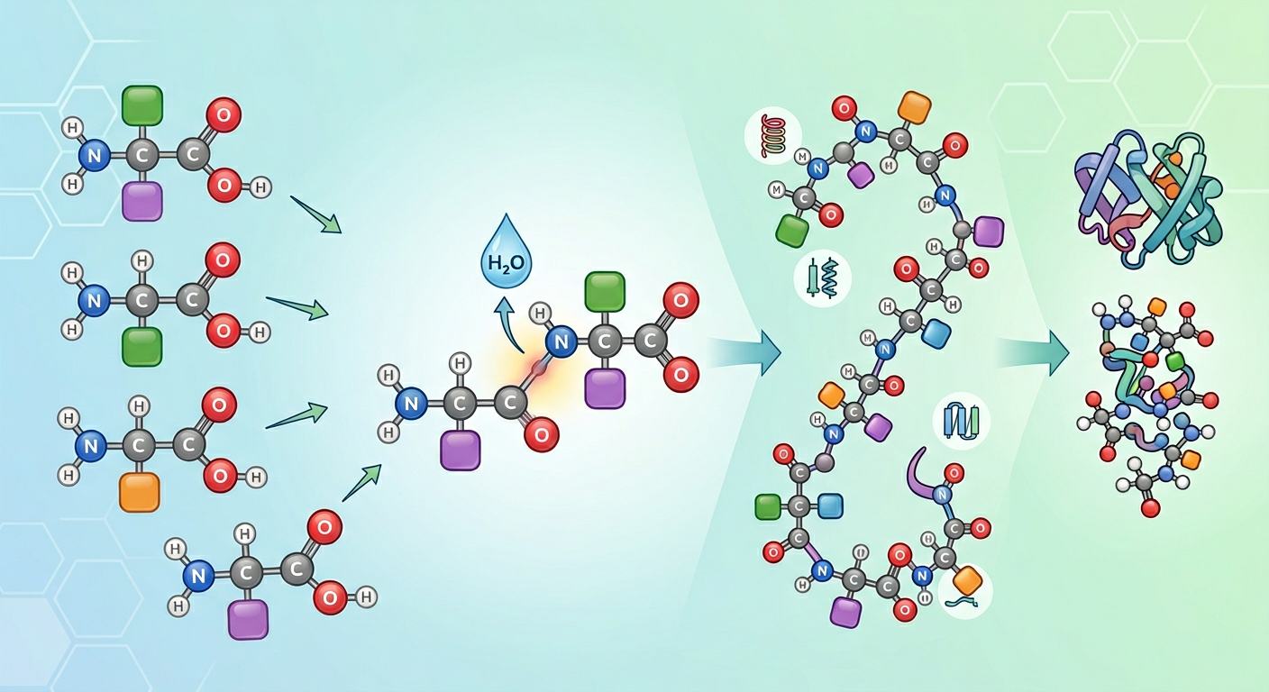
Anti-Aging Peptides: What the Research Actually Shows
Peptides are increasingly marketed for anti-aging purposes. This article examines the evidence base category by category — from collagen peptides to GH secretagogues, GHK-Cu, epithalon, thymosin alpha-1, and MOTS-c — covering mechanisms, what human trials show, and where the evidence ends and extrapolation begins.
Medical Disclaimer: This article is for informational and educational purposes only. It does not constitute medical advice. Anti-aging claims for peptide therapies range from well-supported to speculative. Consult a qualified healthcare provider before beginning any treatment protocol.
“Anti-aging” peptides are everywhere — in skincare serums, supplement stacks, longevity clinic menus, and biohacking forums. The category encompasses everything from widely sold collagen powders to experimental compounds studied for their effects on cellular aging.
The evidence behind these interventions spans a wide spectrum. Some have robust clinical trial data. Others have compelling preclinical data and no human trials. Still others are largely speculative. Understanding where any given compound falls is essential before making decisions about what to put in — or on — your body.
This article examines the science behind each major anti-aging peptide category: the proposed biological mechanisms, what the human and animal evidence actually shows, where independent replication exists, and what the evidence cannot support.
The Biology of Aging: What Are These Peptides Actually Targeting?
Before evaluating evidence, it helps to understand the biological processes of aging that peptide therapies claim to address. A useful framework is the “hallmarks of aging” — the conserved cellular and molecular changes that characterize aged biology across species:
- Genomic instability: accumulating DNA damage over time
- Telomere attrition: progressive shortening of protective chromosome caps
- Epigenetic alterations: changes in gene expression patterns with age
- Loss of proteostasis: declining ability to produce and clear misfolded proteins
- Deregulated nutrient sensing: changes in insulin/IGF-1, mTOR, AMPK, sirtuins
- Mitochondrial dysfunction: declining mitochondrial function and biogenesis
- Cellular senescence: accumulation of non-dividing, pro-inflammatory “zombie cells”
- Stem cell exhaustion: declining tissue regenerative capacity
- Altered intercellular communication: changes in hormonal and inflammatory signaling
Different anti-aging peptides are proposed to address different hallmarks. GH secretagogues primarily target nutrient sensing and body composition. Epithalon targets telomere attrition. GHK-Cu and BPC-157 target tissue repair and altered intercellular communication. MOTS-c targets mitochondrial function. Understanding which hallmark a compound targets helps evaluate whether its proposed mechanism is biologically coherent — and whether the evidence actually shows it working on that target.
Collagen Peptides: The Best-Evidenced Category
The most commercially prevalent anti-aging peptides are collagen peptides — hydrolyzed fragments of collagen protein, typically derived from bovine, porcine, or marine sources and sold as dietary supplements.
The Mechanism: Pro-Hyp Dipeptide Fibroblast Signaling
Collagen is the primary structural protein in skin, connective tissue, tendons, ligaments, cartilage, and bone. It is synthesized by fibroblasts and requires hydroxylated proline residues, which depend on vitamin C as a cofactor.
With age, collagen synthesis declines and collagen cross-linking increases, contributing to skin laxity, wrinkles, joint pain, and bone density loss (Varani et al., 2006). Dermal fibroblasts in aged skin produce less collagen and are less mechanically responsive.
When collagen is hydrolyzed (broken into small fragments for supplementation), the dominant bioactive fragments are dipeptides and tripeptides, particularly Pro-Hyp (proline-hydroxyproline) and related sequences. Research has shown that Pro-Hyp:
- Is absorbed intact across the gut epithelium into circulation
- Accumulates in skin, cartilage, and bone tissue
- Directly stimulates fibroblast proliferation and collagen production in vitro
- Upregulates hyaluronic acid synthesis in skin fibroblasts
This is not simply “you eat collagen, therefore you make more collagen.” The mechanism involves specific bioactive fragments signaling through fibroblast receptors — which is why hydrolyzed collagen peptides show effects that intact collagen does not. The fibroblast receptor pathway has been progressively characterized: Pro-Hyp is absorbed across gut epithelium intact via PEPT1/PEPT2 (SLC15A1/SLC15A2) oligopeptide transporters, which use the electrochemical H⁺ gradient across the brush border membrane to co-transport di/tripeptides against their concentration gradient. Once in circulation, Pro-Hyp reaches dermal fibroblasts where it enters cells through PEPT2 (the higher-affinity isoform expressed in fibroblasts) → intracellular Pro-Hyp activates SP1 (Specificity Protein 1) transcription factor, whose zinc-finger domains recognize GC-rich promoter elements in COL1A1 (α1 chain of type I collagen) and COL1A2 (α2 chain) → increased type I collagen gene transcription. A parallel mTOR arm operates simultaneously: intracellular Pro-Hyp, consistent with amino acid-driven mTOR activation via Rag GTPase/GATOR signaling, stimulates mTORC1/S6K1/4E-BP1 → increased fibroblast protein synthesis rate → faster collagen protein production even without changes in transcription. In hyaluronic acid synthesis, Pro-Hyp also upregulates HAS2 (hyaluronan synthase 2) transcription through the same SP1 axis that drives COL1A1: intracellular Pro-Hyp → SP1 zinc-finger activation → SP1 occupies GC-rich SP1-binding sites in the HAS2 gene proximal promoter (which contains multiple GC-box consensus elements that SP1 recognizes) → HAS2 mRNA transcription; the mTORC1/S6K1 arm additionally increases the intracellular pools of UDP-GlcNAc (by upregulating GFAT1, the rate-limiting enzyme in the hexosamine biosynthetic pathway that generates the GlcNAc substrate) — providing substrate alongside the transcriptional induction — HAS2 polymerizes UDP-glucuronic acid and UDP-N-acetylglucosamine into hyaluronan chains at the inner face of the plasma membrane, extruding the HA polymer directly into the extracellular matrix, accounting for the documented improvement in skin hydration. The combination of transcriptional upregulation (SP1 → COL1A1/COL1A2/HAS2) and translational enhancement (mTORC1 → ribosome biogenesis) at the fibroblast level explains why bioavailable Pro-Hyp dipeptides produce consistent fibroblast responses in vivo that oral intact collagen protein, which is digested to amino acids before absorption, cannot (Shigemura et al., 2009, PMID:19446481).
What Human Trials Show
Skin outcomes: Multiple randomized controlled trials have demonstrated that oral collagen peptide supplementation modestly improves skin elasticity, hydration, and wrinkle depth. Key evidence:
- Proksch et al. 2014 (Skin Pharmacology and Physiology, PMID: 23949208): 69 women, 2.5g or 5g/day for 8 weeks, double-blind placebo-controlled. Significant improvements in skin elasticity, particularly in women over 50.
- Proksch et al. 2014 follow-up (PMID: 24472761): 114 women, 2.5g/day for 8 weeks. Significant improvements in eye wrinkle volume, skin moisture, and skin evaporation.
- A 2019 systematic review of 11 RCTs (PMID: 32436266) found consistent improvements in skin elasticity and hydration across studies, with effect sizes ranging from modest to moderate.
Joint health: Clark et al. 2008 (PMID: 18416885): 147 athletes at Penn State, 10g/day collagen hydrolysate for 24 weeks. Significant reduction in joint pain in athletes during activity. This is one of the better-powered collagen trials.
Bone density: König et al. 2018 (PMID: 29337979): 131 postmenopausal women, specific collagen peptides for 12 months. Significant increase in bone mineral density in the lumbar spine and femoral neck versus placebo, with favorable changes in bone turnover markers. Preliminary but notable finding.
Limitations: Many collagen trials are industry-funded and small. Effect sizes for skin outcomes are real but modest — collagen supplementation is not a substitute for significant sun protection, retinoids, or medical skincare. The bone density finding needs replication in larger trials. These are dietary supplements, not drugs, and aren’t subject to the same regulatory evidence standards.
Practical note: Vitamin C is a required cofactor for collagen hydroxylation — the reason is mechanistic rather than general nutritional. Prolyl-4-hydroxylase (P4H) and the three lysyl hydroxylase isoforms (PLOD1/2/3) are Fe²⁺-dependent, 2-oxoglutarate dioxygenases that hydroxylate proline and lysine residues in nascent procollagen α-chains in the ER lumen. Their catalytic cycle proceeds: the active-site Fe²⁺ binds O₂ → an Fe⁴⁺=O (ferryl) intermediate forms and transfers an oxygen atom to the Pro or Lys side chain → the reaction leaves the iron in the Fe³⁺ state, which cannot initiate the next catalytic cycle. Ascorbate (vitamin C) is the mandatory reductant that recycles Fe³⁺ back to Fe²⁺ after each hydroxylation event. Without sufficient ascorbate, the iron stalls in the Fe³⁺ state → P4H and PLOD activity halts → procollagen Pro and Lys residues remain unhydroxylated → the hydroxyproline hydrogen-bonding network that stabilizes the collagen triple helix (contributing roughly 15 °C to thermal melting temperature) cannot form → misfolded procollagen is retained and degraded in the ER rather than being secreted → net collagen output falls (Myllyharju, 2003, PMID:12837246). This is the precise biochemical basis of scurvy. When supplementing collagen peptides, adequate daily vitamin C (≥75–100 mg in adults) ensures the hydroxylation machinery that incorporates dietary Pro-Hyp into new collagen chains operates at full capacity. Some evidence suggests timing collagen supplementation peri-exercise to exploit the exercise-induced collagen synthesis response. The biological rationale is mechanistically specific and explains why timing matters rather than simply total daily dose. During and immediately after mechanical loading (running, resistance exercise, jump training), tendons and the joint capsule are transiently exposed to tensile and compressive forces that activate Piezo1 (mechanosensitive non-selective cation channel on tenocytes and peri-tendinous fibroblasts) → Ca²⁺ influx → calcineurin activation → NFAT (nuclear factor of activated T-cells) dephosphorylation → NFAT nuclear translocation → drives TGFB1 (TGF-β1) transcription in tenocytes → TGF-β1/ALK5/SMAD2/3 → COL1A1 and TNMD (tenomodulin, a type-II transmembrane protein required for mature tendon fibroblast differentiation) transcriptional upregulation. This exercise-triggered anabolic window peaks approximately 0.5–1 hour after mechanical loading and returns to baseline within 6–8 hours. Shaw et al. (2017, PMID:27852613) demonstrated in a crossover RCT that consuming hydrolyzed collagen + 50mg vitamin C 1 hour before a brief jump-rope exercise bout significantly increased collagen synthesis markers in peri-tendinous tissue compared to the same supplement consumed without exercise or exercise without supplement — establishing that the exercise-induced mechanical signal and the dietary Pro-Hyp substrate are required simultaneously, not independently. The practical implication is that the highest-tissue-concentration window for collagen synthesis — when Piezo1/NFAT/TGF-β1/SMAD2/3 signaling has upregulated the COL1A1 machinery — coincides with the time when absorbed Pro-Hyp dipeptide should be peaking in the circulation, providing substrate for the activated hydroxylation and synthesis enzymes. Vitamin C remains essential during this window: P4H and PLOD activity at the upregulated COL1A1 transcriptional output requires ascorbate as the Fe³⁺→Fe²⁺ electron donor after each hydroxylation cycle (as described in the mechanism section above), meaning the mechanical stimulus can increase collagen gene transcription but cannot convert unhydroxylated procollagen into stable triple-helix without adequate co-factor supply.
Growth Hormone Secretagogues: Targeting the GH Axis
Growth hormone (GH) declines significantly with age — a phenomenon called somatopause. Starting in the late 20s to early 30s, GH pulse amplitude and frequency fall approximately 14% per decade (Iranmanesh et al., 1991). By the 60s, 24-hour GH secretion is roughly 50% of peak young-adult levels.
The functional consequences of somatopause are real but complex: reduced lean muscle mass, increased visceral adiposity, decreased bone density, reduced exercise capacity, and potentially reduced sleep quality. These track with GH/IGF-1 decline.
The GH-Longevity Paradox
Before accepting that “more GH = better aging,” a critical biological nuance: some of the longest-lived animals have reduced GH/IGF-1 signaling. Ames dwarf mice, which are growth hormone-deficient, live up to 50% longer than normal mice (Bartke, 2003, PMID: 12946880). Centenarian humans show enrichment for IGF-1 pathway variants associated with lower IGF-1 activity.
This creates a genuine scientific tension: low GH/IGF-1 may promote longevity at the cellular level through a specific mTOR/autophagy axis, while GH decline in aging is associated with unfavorable body composition and metabolic changes. The mechanism: IGF-1 → IGF-1R → IRS-1 → PI3K → PIP₃ → PDK1 + mTORC2 → Akt → Akt phosphorylates and inhibits TSC2 (tuberous sclerosis complex 2) → TSC2 cannot GAP-inactivate the small GTPase Rheb → GTP-Rheb activates mTORC1 (mechanistic target of rapamycin complex 1). Activated mTORC1 phosphorylates ULK1 at Ser757, directly inhibiting the ULK1 kinase complex — the essential initiator of autophagy (cellular self-digestion that clears damaged organelles, misfolded proteins, and dysfunctional mitochondria) — through two converging mechanisms: (1) AMPK exclusion — AMPK activates ULK1 by phosphorylating it at Ser317 and Ser777; the mTORC1-placed phospho-Ser757 creates a structural clash at the AMPK-docking surface, preventing AMPK from accessing these activating sites — so mTORC1 and AMPK compete directly for ULK1 activation state, with mTORC1 winning when nutrient/IGF-1 signals are high; (2) Pre-initiation complex destabilization — phospho-Ser757 reduces ULK1’s affinity for its obligate co-complex partners ATG13 and FIP200 (RB1CC1), which together with ATG101 form the ULK1/ATG13/FIP200/ATG101 pre-initiation complex required for phagophore nucleation; without stable complex assembly, ULK1 cannot phosphorylate Beclin-1 at Ser15 → Beclin-1 cannot activate the VPS34/PI3K-III lipid kinase complex → insufficient PtdIns3P (PI3P) production at the ER-phagophore assembly site → ATG14 (Barkor) and WIPI2 cannot be recruited via their PI3P-binding domains → the ATG5–ATG12–ATG16L1 E3-ligase complex is not positioned at the nascent autophagosome membrane → LC3-I cannot be lipidated to LC3-II by the ATG7/ATG3 E1/E2 cascade → autophagosome membrane expansion stalls and autophagosomes do not form. When mTORC1 is chronically active (as with sustained high IGF-1), autophagy is continuously suppressed → damaged cellular components accumulate → cellular dysfunction accelerates → aging hallmarks compound faster. In Ames dwarf mice (GH-deficient → very low IGF-1 → reduced PI3K/Akt/mTORC1), ULK1 is constitutively less inhibited → enhanced baseline autophagy → superior cellular housekeeping throughout life → slower accumulation of protein aggregates and dysfunctional mitochondria → the 50% lifespan extension observed. In centenarians, IGF-1 pathway hypomorphic variants appear to preserve this same mTORC1-damping / autophagy-enhancing effect. The tension with somatopause is therefore not a paradox but a dose and timing effect: the question is not “high or low GH/IGF-1 for aging” but rather “what level of mTORC1 activity maintains the autophagy flux needed for cellular quality control while preserving anabolic capacity for lean mass and bone density?”
The resolution is that timing, duration, and magnitude of GH/IGF-1 signaling all matter — and acromegaly represents the most damaging combination of all three, providing the mechanistic basis for why lifelong supraphysiologic GH from early adulthood dramatically shortens lifespan through the same pathways that underlie the longevity paradox. In acromegaly, a pituitary adenoma secretes GH continuously (non-pulsatile) and supraphysiologically, often for a decade or more before diagnosis. First — SOCS3/insulin resistance: non-pulsatile GH means SOCS-3 (induced by each GH/STAT5b pulse) accumulates without the inter-pulse trough needed for proteasomal clearance → progressive JAK2 degradation and IRS-1 Ser307 phosphorylation → peripheral insulin resistance → type 2 diabetes (in ~25% of acromegaly patients); simultaneously GH/JAK2/STAT5b upregulates renal Na-K-ATPase → Na⁺ retention → hypertension (35–50%) → left ventricular hypertrophy and diastolic dysfunction from direct STAT5b/MEF2/ANF-driven cardiomyocyte hypertrophy. Second — constitutive mTORC1/autophagy suppression from youth: lifelong supraphysiologic IGF-1 keeps mTORC1 constitutively active → ULK1 is continuously phosphorylated at Ser757 and inhibited → autophagy flux is suppressed across the entire third, fourth, and fifth decades of life → protein aggregates and dysfunctional mitochondria accumulate from age 20–30 rather than from age 50–60, accelerating all protein-quality-control hallmarks of aging by two to three decades relative to an age-matched eusomatogenic individual. Third — proliferative drive: constitutive IGF-1R/IRS-1/PI3K/Akt/mTOR → CDK4/cyclin D1 in colonic epithelium (via STAT5b-driven suppression of IGFBP-3 → elevated local free IGF-1 → colonocyte PI3K/Akt) → 3–7× increased colorectal cancer risk documented in acromegaly meta-analyses. Therapeutic GH-axis restoration in a GH-deficient adult differs across all three dimensions: it starts from a deficient baseline (not normal), targets the lower end of the physiological range (not above it), restores pulsatility that allows SOCS-3 to clear between pulses (preserving JAK2/IRS-1 function), and produces mTORC1 activation that oscillates with GH pulses — allowing autophagy flux in troughs — rather than constitutive suppression. The goal is not to maximize GH/IGF-1 but to restore the pulsatile, feedback-regulated pattern that maintains the autophagy-anabolism balance that aging disrupts.
The implication for secretagogue use: The goal of GH secretagogue therapy in aging should be restoration toward physiological ranges — not maximization of GH or IGF-1. Secretagogues work by stimulating the pituitary’s own GH release rather than directly injecting GH, which preserves the feedback regulation that prevents runaway elevation.
FDA-Approved Options
Sermorelin (GHRH analog): FDA-approved for GH deficiency in children; used off-label in adults. Has established GH-stimulating effects in published pharmacokinetic and clinical studies. Because sermorelin works within the pituitary’s feedback system, GH elevation is self-limiting — if GH and IGF-1 get too high, the system suppresses release. This is mechanistically distinct from direct GH injection, which bypasses feedback.
Tesamorelin (Egrifta): FDA-approved for HIV-associated lipodystrophy; has demonstrated reduction of visceral adiposity in clinical trials; used off-label in some longevity contexts. The visceral fat selectivity is mechanistically specific — tesamorelin targets the same biology that drives metabolic syndrome and inflammatory aging, for reasons grounded in adipose tissue receptor biology rather than general lipolysis. Tesamorelin is a modified GHRH analogue (trans-3-hexenoic acid conjugation) that activates pituitary GHRH-R to stimulate GH pulses. GH then acts directly on adipocytes via GHR (growth hormone receptor) → JAK2/STAT5b signaling → STAT5b drives transcription of ATGL (adipose triglyceride lipase) and HSL (hormone-sensitive lipase), while simultaneously suppressing LPL (lipoprotein lipase) transcription — promoting triglyceride mobilization and reducing fatty acid uptake from circulating VLDL. The visceral selectivity arises from depot-specific receptor density: visceral omental and mesenteric adipocytes express GHR at approximately 2–3 fold higher density than subcutaneous adipocytes, so an equivalent circulating GH pulse produces disproportionately greater ATGL/HSL induction in the central depot. Additionally, visceral fat has higher portal vein drainage — mobilized FFAs from omental/mesenteric adipocytes enter the hepatic portal directly, where they can be oxidized by the liver rather than redistributed to peripheral depots. The GH/JAK2/STAT5b axis also suppresses IRS-1 Ser307 phosphorylation (the inhibitory modification that accumulates in visceral fat under chronic hyperinsulinemia) in a depot-specific manner — restoring insulin signaling in visceral adipocytes while lipolysis proceeds. This specific visceral adiposity reduction has therapeutic relevance beyond HIV lipodystrophy: visceral fat is the adipose depot most strongly associated with IL-6, TNF-α, and MCP-1 inflammatory cytokine production, adipokine dysregulation, hepatic insulin resistance, and cardiovascular risk — precisely the adipose phenotype of metabolic aging. The clinical tesamorelin data (including a 12-month trial showing significant visceral fat reduction on CT imaging at trunk level) is thus directly mechanistically relevant to the longevity goal of reducing inflammatory adipose burden, even in patients without HIV.
Research Peptides in This Category
Ipamorelin: A selective GHRP (growth hormone releasing peptide) that stimulates GH release through the ghrelin receptor (GHSR-1a). Its selectivity for GHSR-1a over other GPCR targets distinguishes it from earlier generation GHRPs and is the basis of its favorable cortisol/prolactin profile.
Why GHRP-2 and GHRP-6 raise cortisol and prolactin: GHRP-2 and GHRP-6 bind GHSR-1a as primary targets but also have measurable off-target activity at CRF₁/CRF₂ receptors (corticotropin-releasing factor receptors) on hypothalamic neurons and anterior pituitary corticotrophs. CRF receptor activation → Gs/cAMP/PKA in corticotrophs → POMC processing by PC1/3 → ACTH release → adrenal CYP11A1/CYP17A1/CYP21A2/CYP11B1 cascade → cortisol synthesis and secretion. Additionally, GHRP-6 has activity at peptide receptors on hypothalamic dopaminergic neurons that tonically suppress lactotroph prolactin secretion; reducing dopamine output disinhibits lactotrophs → prolactin release. These off-target effects are dose-dependent and are measured in human pharmacokinetic studies as statistically significant cortisol and prolactin elevations after GHRP-2 and GHRP-6 administration.
Why ipamorelin avoids these effects: Ipamorelin’s structural modifications (Aib-His-D-2-Nal-D-Phe-Lys-NH₂ backbone) optimize binding geometry at GHSR-1a with minimal affinity for CRF₁/CRF₂ and no documented activity at hypothalamic dopaminergic receptor subtypes. In the original Raun et al. pharmacological characterization (PMID:9849822), ipamorelin was specifically demonstrated to not elevate ACTH or cortisol at GH-releasing doses — establishing it as the first selective GH secretagogue in its class. This selectivity is clinically relevant for long-term aging protocols where chronic cortisol elevation would counteract the body composition and metabolic goals of GH axis optimization. Commonly combined with CJC-1295 (GHRH analog) to stimulate GH through complementary Gs (CJC-1295/GHRH-R) and Gq (ipamorelin/GHSR-1a) pathways simultaneously — and the mechanistic basis for this synergy is specific enough to explain why the combination produces meaningfully greater GH output than either compound alone at the same individual doses. CJC-1295 (a GHRH analog) binds GHRH-R on pituitary somatotrophs → Gαs → adenylyl cyclase → cAMP elevation → PKA activation → PKA phosphorylates VAMP2 (vesicle-associated membrane protein 2) at Ser75 and Cav1.2/1.3 voltage-gated Ca²⁺ channel regulatory subunits → sensitizes the somatotroph’s L-type voltage-gated Ca²⁺ channels (Cav1.2) to membrane voltage shifts → primes the GH secretory granule exocytosis machinery. The Ca²⁺ that actually triggers exocytosis must come from outside the cell through these L-type channels (following membrane depolarization from the spontaneous somatotroph action potential). Ipamorelin acts through a completely independent Ca²⁺ source: GHSR-1a → Gαq → PLC-β (phospholipase C beta) → cleaves membrane PIP₂ into IP₃ + DAG → IP₃ binds IP₃ receptor (IP₃R) on the ER membrane → ER Ca²⁺ release into the cytoplasm. This ER-released Ca²⁺ and the L-type channel-derived Ca²⁺ from CJC-1295 arm are independently generated from orthogonal sources — the Gq/PLC-β/IP₃/ER pool and the Gs/cAMP/PKA/L-type channel pool — and their cytoplasmic Ca²⁺ contributions are additive at the docking sites of the secretory granule SNARE complex (synaptotagmin/Ca²⁺ sensor + SNAP-25 + synaptobrevin + syntaxin). Because Ca²⁺ sensing by synaptotagmin-I (the GH vesicle Ca²⁺ sensor) requires multiple Ca²⁺ ions (cooperativity coefficient ~4–5), small additive increases in local [Ca²⁺] from two orthogonal sources produce disproportionately large increases in exocytosis rate — explaining why CJC-1295 + ipamorelin combination produces supra-additive GH pulse amplitude at doses where each compound alone would generate only modest responses. Not FDA-approved; compounding status under current rules varies by specific formulation.
CJC-1295 (without DAC): A GHRH analog modified for extended half-life. Has published human pharmacokinetic data showing prolonged GH elevation after administration.
What body composition evidence shows: Studies on GH optimization in GH-deficient adults show improvements in lean mass, reduction of visceral fat, and improved metabolic markers (Johannsson et al., 1997, PMID: 9215274). Studies in healthy older adults without frank GH deficiency show smaller and more inconsistent effects — body composition improvements may occur but effects on functional outcomes are variable. Two mechanistic factors explain the attenuated response compared to GH-deficient populations:
IGF-1 delta magnitude: In GH-deficient adults (the FDA-approved population for tesamorelin/sermorelin), pre-treatment IGF-1 is near-zero. A secretagogue-induced GH pulse elevates IGF-1 from near-zero to the lower range of normal — a large relative increase. Because the IGF-1R/IRS-1/PI3K/Akt/mTORC1 anabolic cascade operates in a dose-responsive range between near-zero and normal IGF-1 concentrations, the absolute increment in Akt Thr308 phosphorylation, mTORC1/S6K1/4E-BP1 signaling, and FOXO1/3a cytoplasmic sequestration is large — producing clinically measurable lean mass accretion and visceral fat reduction as documented in the Johannsson trial. In healthy older adults, baseline IGF-1 sits at the lower end of normal rather than near-zero: mTORC1 and Akt are already partially engaged by tonic basal IGF-1R signaling. A secretagogue-induced increment from the lower end of normal to the mid-normal range produces a proportionally smaller change in downstream anabolic signal magnitude — the body composition response reflects this smaller incremental activation.
Elevated hypothalamic somatostatin tone: Aging is associated with increased hypothalamic somatostatin (SST) secretion — measurable as blunted GH pulse amplitude per unit GHRH stimulus in older versus young adults. SST acts through SST2R (somatostatin receptor 2) on pituitary somatotrophs → Gαi/βγ → adenylyl cyclase inhibition → cAMP falls → PKA activity reduced. This directly attenuates the CJC-1295/GHRH-R Gs/cAMP/PKA arm of somatotroph priming — the same pathway that sensitizes L-type Cav1.2 channels and primes granule exocytosis machinery. Elevated SST tone does not, however, suppress the ipamorelin/GHSR-1a/Gαq/PLC/IP₃/ER-Ca²⁺ arm, because that Ca²⁺ source is orthogonal to cAMP. This is why ipamorelin alone retains some GH-releasing capacity in healthy older adults even under elevated SST tone — but the supra-additive synergy of the CJC-1295 + ipamorelin combination (described above) is partially blunted, because the Gs/cAMP component that normally additively elevates cytoplasmic Ca²⁺ alongside ipamorelin’s ER-Ca²⁺ is antagonized by Gαi. The net GH pulse amplitude per secretagogue dose in healthy older adults is therefore lower than in GH-deficient adults — generating smaller IGF-1 elevations and correspondingly smaller body composition effects, independent of receptor sensitivity.
Safety consideration: Long-term supraphysiologic IGF-1 is associated with increased cancer risk in epidemiological studies. This is the basis for monitoring IGF-1 levels and targeting physiological rather than supraphysiologic ranges. Baseline and follow-up IGF-1 testing is required for responsible GH secretagogue protocols.
GHK-Cu (Copper Peptide): Skin and Tissue Repair
GHK-Cu (glycine-histidine-lysine copper complex) is a naturally occurring tripeptide that binds copper and is present in human plasma, saliva, and urine. It was first isolated by Loren Pickart in the 1970s and has one of the longer research histories of any cosmetic/therapeutic peptide.
The Mechanism: Gene Expression Remodeling
The most striking finding in GHK-Cu research is the breadth of gene expression changes it induces. A 2012-2014 analysis by Pickart and colleagues, and a 2018 re-analysis of the GEO gene expression database (PMID: 29988121), found that GHK-Cu regulates over 4,000 human genes, including:
- Upregulation of collagen, elastin, and glycosaminoglycan synthesis genes
- Modulation of inflammation-related genes
- Upregulation of antioxidant and DNA repair genes
- Downregulation of genes associated with cancer metastasis in some analyses
Wound healing: GHK-Cu has documented wound healing effects in animal models and some human studies. It promotes fibroblast migration, collagen synthesis, and angiogenesis through mechanisms distinct from BPC-157. The angiogenic pathway begins with GHK-Cu’s Cu²⁺ chelate: copper is an obligate cofactor for lysyl oxidase (LOX) — the enzyme that cross-links collagen and elastin fibrils by oxidizing lysine side chains to allysine aldehydes for subsequent Schiff base formation — and for copper,zinc-superoxide dismutase (SOD1/CuZnSOD), which dismutates superoxide (O₂•⁻) to H₂O₂. Controlled intracellular H₂O₂ generated by SOD1 acts as a second messenger that oxidizes the catalytic Fe²⁺ in prolyl hydroxylase domain (PHD) proteins (PHD1/2/3 — the oxygen-sensing HIF-1α hydroxylases) — iron oxidation inhibits PHD activity, preventing them from hydroxylating the prolyl residues on HIF-1α that trigger its VHL-mediated ubiquitination and proteasomal degradation. The result is HIF-1α stabilization in normoxic conditions: non-degraded HIF-1α translocates to the nucleus → dimerizes with ARNT/HIF-1β → the HIF-1α/ARNT heterodimer binds HREs (hypoxia response elements) in the proximal promoters of VEGF-A, VEGFR1, and SDF-1 → VEGF-A transcription. Secreted VEGF-A then binds VEGFR2 (KDR) on endothelial cells → VEGFR2 kinase domain trans-autophosphorylation at Tyr1175 → PLCγ/IP₃/DAG → PKC-dependent RAF-MEK-ERK1/2 → endothelial cell proliferation; Tyr1214 phosphorylation → Cdc42/p38-MAPK → endothelial cell migration (filopodial extension by tip cells). A parallel arm operates through TGF-β upregulation: GHK-Cu induces TGF-β1 secretion from fibroblasts → TGF-β1 binds the TGF-βRII/TGF-βRI (ALK5) heterodimeric receptor complex → ALK5 phosphorylates SMAD2 Ser465/467 and SMAD3 Ser423/425 → SMAD2/3 release from SARA (SMAD Anchor for Receptor Activation) → SMAD2/3 bind SMAD4 → nuclear complex → COL1A1, COL1A2, FN1 (fibronectin), and CTGF (connective tissue growth factor) gene transcription → fibroblast ECM deposition. LOX then cross-links the deposited collagen, restoring tensile strength. This combination of HIF-1α/VEGF-driven neovascularization (supplying oxygen and nutrients) with SMAD2/3-driven ECM rebuilding (structural remodeling) explains why GHK-Cu wound healing includes both angiogenesis and matrix deposition phases — operating through copper biochemistry rather than VEGFR2 direct binding (as in BPC-157) (PMID:29988121).
Skin evidence: Topical GHK-Cu at 1-4% concentrations has been studied in several controlled trials showing improvements in skin thickness, wrinkle depth, and firmness. A 1993 Leyden et al. study in 67 subjects demonstrated improvements in fine lines and overall skin appearance comparable to retinoic acid at 3 months — though this finding has not been extensively replicated in large modern trials.
The systemic vs. topical distinction: GHK-Cu is well-characterized as a topical cosmetic ingredient with meaningful penetration into the dermis. Systemic administration (injectable) is a separate and much less characterized use case, with essentially no human clinical trial data for systemic GHK-Cu protocols.
Regulatory status: Topical GHK-Cu products are widely available as cosmetics. Systemic GHK-Cu compounding exists but is at the research frontier with limited human evidence.
Epithalon: Telomere Claims and Reality
Epithalon (Ala-Glu-Asp-Gly, also written “Epitalon”) is a synthetic tetrapeptide corresponding to a sequence from epithalamin, a natural pineal gland polypeptide. It has attracted the most attention in longevity circles due to claims about telomere lengthening.
The Telomere Biology Context
Telomeres are repetitive DNA sequences at the ends of chromosomes that protect against genomic instability. With each cell division, telomeres shorten. When they become critically short, cells enter senescence or apoptosis. Telomere attrition is one of the “hallmarks of aging” — associated with aging-related diseases and shorter lifespan in epidemiological studies.
Telomerase is the enzyme that can lengthen telomeres by adding repetitive sequences. It is active in stem cells and cancer cells but downregulated in most somatic cells. Activating telomerase in normal somatic cells is an appealing longevity strategy in theory — but with a significant caveat: cancer cells extensively upregulate telomerase to achieve replicative immortality. Any telomerase activator raises cancer promotion concerns that are mechanistically specific, not merely theoretical. The risk operates through two distinct molecular dimensions simultaneously. The first is relief of the replicative senescence tumor suppression barrier: throughout life, rare somatic cells acquire pre-cancerous oncogenic mutations — activating mutations in RAS/KRAS/BRAF, or loss-of-function mutations in p53 (TP53) or RB1. In cells with normal telomere attrition, progressive telomere shortening eventually triggers the ATM/ATR → p53 → p21 → G1 arrest senescence program that permanently removes these mutation-harboring cells from the replicating pool before they can accumulate additional hits toward malignancy. Telomerase restoration in these cells removes this replicative barrier: the oncogene-harboring cell that would have senesced upon telomere crisis instead continues dividing, accumulates additional mutations, and now has both an oncogenic driver and extended replicative lifespan — a pre-malignant combination. The second dimension is non-canonical TERT transcriptional co-activation functions independent of telomere elongation: TERT protein is not solely a reverse transcriptase extending telomeres — it has well-characterized roles as a transcriptional co-activator of two of the most common oncogenic pathways. TERT translocates to the nucleus in complex with β-catenin and directly co-activates Wnt/β-catenin/TCF target gene transcription (including CCND1/Cyclin D1, MYC, and AXIN2), the same Wnt program that is the canonical driver in colorectal and other cancers. TERT also directly interacts with the MYC/MAX complex at MYC target gene E-box elements, enhancing transcription of ribosomal biogenesis factors, metabolic enzymes, and cell-cycle accelerators (CDK4, CCND1) that support rapid proliferation. Importantly, these non-canonical TERT functions are fully active in cells where TERT is exogenously induced before telomere crisis — meaning telomerase activation doesn’t simply provide a replicative lifespan extension but concurrently upregulates two major oncogenic transcriptional programs in any cell that expresses it. Cancer cells exploit exactly these non-canonical TERT activities for their proliferative and metabolic advantages, in addition to telomere maintenance. The theoretical concern for longevity telomerase activators is therefore the combination of removing the senescence barrier (allowing pre-malignant cells to persist) and simultaneously activating Wnt/β-catenin and MYC programs (providing proliferative drive) in those cells — neither alone is sufficient for malignancy, but together they markedly accelerate progression through the remaining mutation landscape.
What Epithalon Research Shows
The primary research base comes from Vladimir Khavinson and colleagues at the St. Petersburg Institute of Bioregulation and Gerontology. Key claims:
- In cell culture studies, epithalon activated telomerase (specifically TERT, the catalytic subunit) in human fetal fibroblasts and extended telomere length (Khavinson et al., 2003, PMID: 14583750). The proposed mechanism involves epigenetic remodeling of the TERT gene promoter. In most somatic cells, TERT transcription is silenced by a combination of CpG methylation at the TERT promoter’s GC-rich core and histone deacetylation-driven chromatin compaction — these modifications block SP1 and c-Myc/Max from accessing their cognate binding sites within the TERT proximal promoter and E-box elements, keeping the TERT gene transcriptionally inaccessible. Epithalon (AEDG) is proposed to interact with chromatin-associated regulatory proteins and promote histone H3/H4 acetylation at the TERT promoter → chromatin decompaction → SP1 and c-Myc access → TERT mRNA transcription → TERT protein assembles with the RNA component TERC (telomerase RNA component, which provides the CCCCAA template strand for TTAGGG synthesis) and the stabilizing pseudouridylase/assembly complex (dyskerin/DKC1, NOP10, NHP2) → functional telomerase holoenzyme → reverse transcribes TTAGGG repeats onto chromosome 3’ G-strand overhangs, extending telomere length at each replication. Critically, the specific chromatin interaction partner for the AEDG tetrapeptide has not been identified in published research — the epigenetic mechanism is inferred from downstream gene expression and telomerase activity measurements, not from direct molecular binding studies characterizing what AEDG physically contacts in chromatin. This mechanistic uncertainty is important context: TERT activation was demonstrated (mRNA quantification, TRAP telomerase activity assays), but the proximal molecular trigger remains uncharacterized, which is a significant gap in the mechanistic evidence chain
- In various animal studies (mice, rats, fruit flies), epithalon treatment was associated with extended lifespan and reduced age-related pathology
- In small human studies — primarily elderly patients in Russian clinical settings — epithalon was reported to improve immune function, retinal function, and some hormonal parameters
Critical scientific limitations:
-
Geographic and independent replication concentration: The vast majority of epithalon research comes from a single Russian research group. Independent replication by groups in other countries is extremely limited. This is similar to the BPC-157 literature’s Sikiric lab concentration problem.
-
Study design quality: Published human studies are generally small, non-randomized, and without adequate blinding or placebo control. None meet modern RCT standards.
-
Telomere biology complexity: Telomere lengthening in cell culture does not prove lifespan extension in vivo. The relationship between average telomere length, individual cell senescence, and organismal aging is not linear — and the specific molecular reasons for this non-linearity determine which telomere measurements are and are not meaningful. Three independent mechanisms explain why epithalon’s documented increase in mean telomere length is insufficient to infer senescence protection or lifespan extension. First, the shortest-telomere rule: senescence is not triggered by average telomere length but by the shortest telomere in any given cell. When even a single chromosome end shortens below approximately 3 kb of TTAGGG repeats, the exposed 3’ G-overhang is recognized as double-strand DNA damage by the MRN complex (MRE11–RAD50–NBS1) → ATM recruitment and autophosphorylation at Ser1981 → ATM phosphorylates CHK2 at Thr68 → CHK2 phosphorylates p53 at Ser20 → MDM2-mediated p53 ubiquitination is blocked → p53 accumulates → transcription of p21/CDKN1A → CDK2/CDK4 inhibition → permanent cell cycle arrest and senescence. A cell whose average telomere rises from 4 kb to 6 kb through exogenous TERT activation may still harbor a single 1.8 kb telomere that triggers ATM/p53/p21 arrest; mean elongation is irrelevant if the shortest telomere remains below the crisis threshold. Second, tissue-specific cell type sensitivity: post-mitotic neurons do not replicate and therefore do not shorten telomeres through replicative attrition regardless of telomerase status — neuronal TERT activation is irrelevant to neurodegeneration. Hematopoietic stem cells (HSCs) divide frequently and are exquisitely sensitive to telomere shortening; this is why telomerase deficiency diseases (dyskeratosis congenita, caused by DKC1 mutations reducing TERC stability or TERT mutations) manifest in rapidly-renewing tissues (bone marrow aplasia, gut epithelial failure, mucocutaneous abnormalities) rather than in post-mitotic brain. A systemic telomere measurement — typically from peripheral blood leukocytes — reflects mainly lymphocyte and granulocyte telomere length, not the HSC compartment or intestinal crypt stem cells that are functionally rate-limited by shortening. Epithalon’s TRAP-assay telomerase readout in fetal fibroblasts neither characterizes nor predicts what happens in the stem cell compartments most relevant to human aging tissue maintenance. Third, telomere length heritability: inter-individual variation in telomere length at birth spans 5–15 kb, with heritability estimated at 70–80%. This constitutional variation dwarfs the ~0.5–1 kb per decade of replicative attrition, meaning individuals with constitutively short starting lengths reach senescence-inducing thresholds decades earlier than individuals with constitutively long starting lengths — independent of any intervention, and independent of average-population telomere aging rates. Associations between telomere length and longevity in population studies reflect this heritable distribution rather than proving that extending any individual’s average telomere length produces proportional lifespan benefit. The combined implication of these three mechanisms: an experiment demonstrating that AEDG increases mean TTAGGG abundance in a fibroblast culture (measured by TRAP activity reflecting elongated mean telomeres) does not establish that epithalon would (a) elongate the shortest telomere in a senescent-cell-at-risk population, (b) act in the correct cell compartments, or (c) move individuals from a short-heritability to a long-heritability functional outcome.
-
Cancer risk question: No long-term safety data exists. The theoretical concern about telomerase activation and cancer promotion has not been addressed by the published research.
Bottom line: Epithalon has a mechanistically interesting profile and some preliminary human data, but the evidence quality is significantly below what would be needed to recommend clinical use. It is not FDA-approved, not eligible for US compounding pharmacy dispensing, and available only through gray-market channels with associated quality risks.
Thymosin Alpha-1: Immune Function in Aging
Thymosin Alpha-1 (Tα1) is a 28-amino acid peptide naturally produced by thymic epithelial cells. It is an endogenous regulator of T-cell maturation and immune function.
The Immunosenescence Context
The thymus — the organ that trains T cells — progressively involutes (shrinks) with age. By age 40, the thymus is roughly 50% replaced by adipose tissue; by age 70, it produces minimal new T cells. The molecular mechanism driving this involution converges on FOXN1 (Forkhead box N1), the master transcription factor for thymic epithelial cell (TEC) identity. FOXN1 maintains TEC expression of DLL4 (the Notch ligand required to commit T-cell precursors to the T-cell lineage), MHC-II, and AIRE (autoimmune regulator), which together create the three-dimensional epithelial scaffold required for positive and negative T-cell selection. FOXN1 expression declines in aged TECs through two converging inputs. First, Wnt signaling atrophy: developing thymocytes secrete Wnt3a, which normally signals through Frizzled/LRP5-6 receptors on adjacent TECs → Dvl recruits Axin to the membrane, disrupting the APC/Axin/CK1α/GSK-3β destruction complex → β-catenin escapes Ser33/37 phosphorylation and β-TrCP-mediated ubiquitination → nuclear β-catenin/TCF drives FOXN1 transcription; as thymocyte output falls with age, Wnt3a supply weakens, the destruction complex re-engages, β-catenin is degraded, and FOXN1 transcription declines. Second, the IGF-1/Akt/FOXO1 adipogenic axis: somatopause-driven IGF-1 decline → reduced IGF-1R/IRS-1/PI3K/Akt signaling in TECs → less Akt-mediated FOXO1 Ser256 phosphorylation → FOXO1 nuclear accumulation in TECs → FOXO1 drives transcription of C/EBPα and PPARγ, the master adipogenic differentiation factors → TECs undergo lipid droplet accumulation, lose epithelial junction proteins, and lose FOXN1 expression — a TEC-to-adipocyte transdifferentiation that replaces functional thymic stroma with metabolically inert adipose-like cells, explaining the ~50% adipose content by age 40. Androgen receptor signaling accelerates this process: AR is expressed in thymic stromal cells and directly suppresses FOXN1 transcription; gonadectomy reverses thymic involution in animal models, and androgen-ablation therapy in men with prostate cancer transiently restores naïve T-cell output — linking the sex steroid decline of aging to faster thymic collapse. This thymic involution drives immunosenescence: the age-related decline in immune function characterized by:
- Reduced naïve T cell output (fewer new T cells trained for novel pathogens)
- Accumulation of exhausted or senescent memory T cells
- Chronic low-grade inflammation (“inflammaging”)
- Reduced vaccine responsiveness in older adults
- Increased susceptibility to infections and cancer
Thymosin Alpha-1’s proposed role in aging is to compensate partially for this thymic decline by supporting T-cell maturation through its signaling effects on dendritic cells and T-cell precursors. The molecular mechanism — more fully characterized than most immune-modulating peptides in this category — operates primarily through a TLR/DC/IL-12/STAT4 axis.
The TLR-DC activation arm. Tα1 engages Toll-like receptors on plasmacytoid and myeloid dendritic cells, with strongest evidence for TLR9 (which recognizes unmethylated CpG motifs, a pattern found in bacterial and viral DNA, suggesting Tα1 mimics a danger signal to prime the innate-adaptive interface). TLR9 engagement recruits the adaptor protein MyD88 → MyD88 assembles the IRAK4/IRAK1 complex (IL-1 receptor-associated kinases) → IRAK4 autophosphorylates and phosphorylates IRAK1 → IRAK1 dissociates from MyD88 → TRAF6 (TNF receptor-associated factor 6) → TRAF6 undergoes K63-linked autoubiquitination → recruits TAK1/TAB1/TAB2 complex → TAK1 phosphorylates IKKβ → IKKβ phosphorylates IκBα at Ser32/Ser36 → IκBα undergoes K48 ubiquitination and proteasomal degradation → NF-κB p65/p50 heterodimer is released from cytoplasmic sequestration → nuclear translocation → NF-κB binds κB response elements in the IL-12A (p35 subunit) and IL-12B (p40 subunit) gene promoters → transcription and secretion of IL-12 p70 (the biologically active heterodimer) by activated dendritic cells (Romani et al., 2004, PMID:15148335).
The IL-12/STAT4/T-bet Th1 differentiation arm. DC-secreted IL-12 p70 engages the IL-12Rβ1/IL-12Rβ2 heterodimeric receptor on naïve CD4⁺ T cells (and CD8⁺ cytotoxic precursors). Receptor engagement → JAK2 (constitutively associated with IL-12Rβ1) and TYK2 (with IL-12Rβ2) transphosphorylate each other → JAK2/TYK2 phosphorylate STAT4 at Tyr693 → STAT4 homodimerization → nuclear translocation → STAT4 binds serum response elements (SREs) in the IFN-γ (IFNG) promoter and the T-bet (TBX21) promoter → dual transcription of IFN-γ (the canonical Th1 effector cytokine, driving macrophage activation, viral clearance, and antitumor surveillance) and T-bet (the master Th1 transcription factor). T-bet then locks in Th1 identity through three parallel mechanisms: (1) T-bet transactivates IL-18Rα expression → enables IL-18 co-stimulation for sustained IFN-γ production in tissue; (2) T-bet upregulates CXCR3 → chemotaxis toward CXCL10/IP-10 gradients in inflamed tissues, enabling T cell homing to sites of viral infection or tumor; (3) T-bet binds and functionally represses GATA-3 (the master Th2 transcription factor) through protein-protein interaction — ensuring that Tα1-driven T cells do not produce IL-4 or IL-5 but maintain Th1 effector programs (Szabo et al., 2000, PMID:10707990). The net result is a DC→IL-12→STAT4→T-bet cascade that reconstitutes Th1 surveillance capacity even when the thymic output of naïve T cells is reduced — partially compensating for immunosenescence at the T-cell functional level rather than the thymic production level.
Human Evidence
Tα1 (brand name Zadaxin, approved in several countries) has the most meaningful human clinical evidence of any compound in the immune aging category:
- Hepatitis B/C: Multiple trials across Asia and Europe showing improved response to antiviral treatment when Tα1 is added
- Cancer adjuvant: Studies showing improved immune function in oncology patients receiving chemotherapy; some evidence for improved survival in specific cancers
- COVID-19: A 2024 RCT (PMID: 41887933) in severe liver failure patients with COVID-19 showed benefit in the Tα1 group
- Vaccine response: Some evidence for improved response to influenza and hepatitis B vaccines in elderly subjects. The mechanistic basis involves compensating for age-related dendritic cell (DC) dysfunction. In immunosenescent DCs, TLR9 surface expression falls progressively because aging monocytes and DCs upregulate miR-146a, a microRNA that targets IRAK1 and TRAF6 mRNAs — both essential adapter proteins in the MyD88/IRAK4/TRAF6 signaling cascade downstream of TLR9. When IRAK1 and TRAF6 protein are depleted, each TLR9 engagement generates attenuated NF-κB activation and consequently reduced IL-12 p70 secretion per DC, impairing antigen-specific Th1 priming. Tα1 functions as a TLR9 agonist that amplifies residual TLR9/MyD88/IRAK4 signaling within this already-attenuated system: even with reduced IRAK1/TRAF6 protein, sufficient NF-κB nuclear translocation and IL-12 p70 production can be achieved when TLR9 occupancy is maximized by exogenous agonism. The resulting IL-12 p70 drives STAT4 phosphorylation (Tyr693) → T-bet transcription factor expression → IFN-γ-producing antigen-specific Th1 CD4+ cells → CD8+ cytotoxic T lymphocyte priming and T-follicular helper–dependent IgG class switching to high-affinity vaccine-specific antibodies. This mechanism explains why Tα1 benefit in vaccine response trials is most pronounced in elderly subjects (where the miR-146a/IRAK1/TRAF6 deficit is greatest) rather than in young adults with intact TLR9 signaling
For general “immune aging” use in otherwise healthy older adults: The evidence is more limited. The disease-context studies demonstrate biological activity and reasonable safety; they don’t directly validate routine use for healthy older people seeking immune optimization.
Tα1 is compoundable through some US facilities and is FDA-approved in other countries. It represents one of the more clinically credible compounds in the research peptide space due to its existing human evidence base in disease populations.
MOTS-c: The Mitochondrial Peptide
MOTS-c (Mitochondrial Open Reading Frame of the 12S rRNA-c) is a 16-amino acid peptide encoded in mitochondrial DNA — making it unusual among peptides, which are typically encoded in nuclear DNA.
The Mechanism
MOTS-c regulates AMPK (AMP-activated protein kinase), a key cellular energy sensor. What distinguishes MOTS-c from other AMPK activators is how it activates AMPK — through an unusual nuclear mechanism involving the folate metabolic cycle rather than direct sensing of the AMP:ATP ratio.
MOTS-c is a 16-amino acid peptide (MRWQEMGYIFYPRKLR) encoded within the 12S rRNA gene of mitochondrial DNA. Under metabolic stress, MOTS-c is released from mitochondria and translocates directly into the nucleus — an unusual destination for a mitochondrially-encoded signaling peptide. In the nucleus, MOTS-c interferes with two enzymes of the folate one-carbon metabolic cycle: ATIC (AICAR transformylase, bifunctional) and MTHFS (methenyltetrahydrofolate synthetase). These enzymes are required for converting AICAR (5-aminoimidazole-4-carboxamide ribonucleotide) onward toward IMP (inosine 5’-monophosphate) in de novo purine biosynthesis. When MOTS-c inhibits ATIC and MTHFS, AICAR accumulates intracellularly. AICAR is a well-characterized AMP mimetic — it binds to the regulatory γ-subunit of AMPK at the same allosteric site as AMP, triggering the same conformational activation as if the cellular energy charge had fallen (AMP:ATP ratio risen). This indirect AMPK activation mimics the metabolic signal of energy depletion without requiring actual ATP expenditure (Lee et al., 2015, PMID:25738459).
AMPK activation then drives the downstream cascade:
- Phosphorylates and inhibits ACC (acetyl-CoA carboxylase) → reduces malonyl-CoA → disinhibits CPT1 → enhanced mitochondrial fatty acid import and β-oxidation
- Activates TSC2 and inhibits Raptor → mTORC1 suppression → ULK1 activation → autophagy initiation (selective clearance of damaged mitochondria via mitophagy)
- Elevates intracellular NAD⁺ (by suppressing ATP-consuming anabolic pathways) → activates SIRT1 → SIRT1 deacetylates PGC-1α → PGC-1α drives transcription of mitochondrial biogenesis and oxidative phosphorylation genes
- Promotes GLUT4 translocation to the plasma membrane in muscle and adipose tissue → improved insulin-independent glucose uptake. The molecular mechanism: AMPK directly phosphorylates AS160 (TBC1D4, TBC1 domain family member 4) at Ser588 — AS160 is a Rab-GTPase-activating protein (Rab-GAP) that normally maintains Rab8A, Rab10, and Rab14 in their inactive GDP-loaded states by hydrolyzing their GTP, preventing GLUT4 storage vesicles from docking at the plasma membrane. AMPK-driven Ser588 phosphorylation inactivates AS160’s GAP catalytic domain → Rab8A/10/14 remain GTP-loaded → GTP-Rab recruits ELKS/RIMS-like vesicle tethering effectors and facilitates assembly of the SNARE fusion complex (VAMP2 on the GLUT4 vesicle membrane + SNAP-23 + syntaxin-4 on the target plasma membrane) → GLUT4 storage vesicle docks and fuses → GLUT4 transporter surface density increases → facilitated glucose uptake proceeds. The critical insulin-independence operates through distinct phosphosite architecture: the AMPK-specific AS160 Ser588 site is structurally separate from the insulin/Akt-driven AS160 Thr642 phosphosite — two independent molecular switches that inactivate the same Rab-GAP via different kinases. In aged, insulin-resistant skeletal muscle where the PI3K/Akt pathway is blunted by IRS-1 Ser307 inhibitory phosphorylation from chronic low-grade inflammation (the same inflammaging that MOTS-c’s AMPK cascade attenuates via mTORC1 suppression), the Akt-dependent Thr642 switch is impaired — but AMPK-dependent Ser588 phosphorylation operates independently of this blockade, explaining why glucose disposal is preserved in MOTS-c-treated insulin-resistant aged animals even when insulin/Akt responsiveness is reduced
Circulating MOTS-c levels decline with age in humans, and higher MOTS-c levels have been associated with longevity in some centenarian studies — suggesting it may be part of the endogenous signaling that supports healthy aging. The mechanistic basis for this age-related decline is rooted in mitochondrial DNA heteroplasmy accumulation: as cells divide and mitochondria replicate over decades, replication errors and oxidative damage to mitochondrial DNA accumulate as heteroplasmic mutations — variant copies co-existing with wild-type copies within the same cell. The mitochondrial 12S rRNA gene locus where MOTS-c is encoded is subject to this heteroplasmy drift; as the proportion of mutant 12S rRNA copies increases, TFAM (mitochondrial transcription factor A) — which must bind the D-loop to initiate 12S rRNA transcription — produces progressively less functional 12S rRNA transcript. Since MOTS-c is translated from this same transcript, total cellular MOTS-c production falls in parallel with heteroplasmy accumulation. The downstream consequence follows the AICAR/AMPK cascade in reverse: less MOTS-c → less ATIC/MTHFS inhibition in the nucleus → AICAR does not accumulate → AMPK is not allosterically activated by the AICAR/AMP-mimetic route → mTORC1 is progressively less suppressed (less TSC2 activation by AMPK, less direct Raptor phosphorylation) → ULK1 phosphorylation at Ser317/Ser777 declines → autophagy flux slows → damaged mitochondria that would have been cleared by AMPK-driven mitophagy now accumulate → a feed-forward cycle in which declining mitochondrial quality produces more heteroplasmy, which produces less MOTS-c, which produces less mitophagy-driven quality control. Centenarian-associated genetic variants near the mitochondrial 12S rRNA locus or in nuclear-encoded mitochondrial maintenance genes (POLG, TWNK helicase) that reduce somatic heteroplasmy accumulation rate would, by this mechanism, maintain higher MOTS-c production, preserve AMPK activation efficiency, and sustain mTORC1-regulated proteostasis into very old age — making MOTS-c a readout of mitochondrial integrity rather than an independent longevity effector per se. Exercise increases circulating MOTS-c levels, which may partly explain how exercise produces systemic metabolic benefits even in non-exercising tissues.
Evidence Status
MOTS-c research is at an earlier stage than most compounds discussed here:
- Animal studies show improvements in metabolic function, exercise capacity, and insulin sensitivity — and the specific endpoints map directly to the AMPK downstream cascade described above. Improved metabolic function (reduced fat mass, improved lipid profiles in MOTS-c-treated mice) reflects ACC phosphorylation → malonyl-CoA suppression → CPT1 disinhibition → enhanced skeletal muscle and hepatic mitochondrial fatty acid β-oxidation. Improved insulin sensitivity corresponds to GLUT4 translocation via AMPK’s direct phosphorylation of AS160/TBC1D4 at Ser588 — an Akt-independent GLUT4 trafficking signal that operates even in states of reduced insulin receptor activation, explaining why the effect is observed in aging animals where Akt responsiveness is blunted. Improved exercise capacity maps to SIRT1 deacetylation of PGC-1α → mitochondrial biogenesis in skeletal muscle → higher oxidative phosphorylation capacity per fiber → increased VO₂max. MOTS-c also reduced age-associated frailty and adiposity in aged female mice, with effects proportional to the magnitude of the AICAR/AMPK activation produced — consistent with the ATIC/MTHFS/folate-cycle mechanism rather than a non-specific peptide effect (Kim et al., 2021, PMID:31011227)
- Preliminary human data suggests age-related MOTS-c decline and possible associations with healthspan markers. In centenarian cohort analyses, individuals carrying genetic variants associated with higher mitochondrial 12S rRNA transcription output (and thus higher MOTS-c expression) show metabolic profiles consistent with better-preserved insulin sensitivity and lower visceral adiposity at advanced age — suggesting endogenous MOTS-c level may be a component of the metabolic resilience phenotype associated with exceptional longevity, though the sample sizes are small and confounding is not fully characterized
- No published RCTs in humans; no clinical pharmacokinetic data established
The mechanism underlying age-related MOTS-c decline is now partially understood and connects directly to the same mitochondrial dysfunction that MOTS-c itself counteracts. MOTS-c is encoded within the 12S rRNA gene of the mitochondrial genome — not the nuclear genome — so its production depends on the transcriptional integrity of mtDNA rather than nuclear gene-expression machinery. With aging, somatic mtDNA point mutations and deletions accumulate through replication error and oxidative damage; the proportion of cells carrying mutant mtDNA alongside wild-type copies (heteroplasmy) increases progressively in post-mitotic tissues including skeletal muscle, liver, and adipose. Beyond a tissue-specific threshold, elevated heteroplasmy impairs mitochondrial transcription factor A (TFAM)-driven mtDNA transcription, reducing steady-state levels of all mitochondrially-encoded transcripts including the 12S rRNA precursor from which MOTS-c is translated. Separately, the age-related decline in NAD⁺ reduces SIRT1 and SIRT3 deacetylase activity; TFAM becomes hyperacetylated and binds mtDNA with reduced affinity, further suppressing 12S rRNA transcription — the structural basis being that TFAM contains two HMG-box (high-mobility group box) DNA-binding domains that contact the mtDNA minor groove and introduce a ~180° bend required for stable transcription initiation complex formation at the LSP (light strand promoter) and HSP1 (heavy strand promoter 1) within the D-loop control region; the minor-groove binding interface is dominated by positively charged lysine residues whose electrostatic attraction to the negatively charged mtDNA phosphate backbone anchors TFAM and establishes the bending angle. GCN5L1 (a mitochondrially-localized acetyltransferase) constitutively acetylates TFAM at lysines within and flanking the HMG-box binding interface (K119, K146, K157 as resolved by mass spectrometry); SIRT3 (the primary mitochondrial sirtuin deacetylase) normally reverses this modification using NAD⁺ as cofactor. When NAD⁺ falls with age, SIRT3 catalytic activity declines → GCN5L1-driven acetylation of TFAM accumulates without reversal → the neutralized lysine ε-amino groups lose their positive charge → electrostatic attraction to the mtDNA phosphate backbone is weakened → TFAM cannot achieve the D-loop bending angle required for transcription factor assembly → TFAM binds mtDNA with reduced affinity and shorter dwell time (PMID:30890426). This dual suppression — heteroplasmy-driven transcription impairment plus NAD⁺/SIRT1/TFAM axis dysfunction — explains why circulating MOTS-c falls progressively after the fourth decade and why exercise partially reverses it: acute exercise activates PGC-1α/NRF1/TFAM-driven mitochondrial biogenesis → transient increases in mtDNA copy number and transcriptional output → MOTS-c burst → nuclear AMPK activation. The same age-related mitochondrial dysfunction that reduces basal MOTS-c also blunts the PGC-1α biogenesis response to a given exercise stimulus, creating a self-reinforcing deficit that worsens as mitochondrial quality declines.
MOTS-c is primarily of research interest at this stage. Not available through US compounding pharmacies.
Cellular Senescence and FOXO4-DRI: An Emerging Peptide Target
Of all the hallmarks of aging listed above, cellular senescence is perhaps the most amenable to targeted intervention — and has produced some of the most striking animal research in the longevity peptide field. A peptide specifically designed to eliminate senescent cells (FOXO4-DRI) represents a different conceptual approach from the compounds discussed elsewhere in this guide: rather than supplementing a declining signal or slowing a process, it removes a class of dysfunctional cells.
The Biology of Senescent Cells and SASP
Senescent cells are cells that have permanently exited the cell cycle — they stop dividing but do not die. They accumulate with age due to replicative exhaustion (cells that have divided too many times and reached telomere length limits), unrepairable DNA damage triggering permanent cell-cycle arrest, and oncogene activation in pre-cancerous cells that senesce as a tumor-suppression mechanism rather than becoming malignant.
In a young organism, senescent cells are efficiently cleared by the immune system — primarily by NK cells and CD8⁺ cytotoxic T cells. With age, immune surveillance of senescent cells declines through three mechanistically distinct failure modes, and senescent cells accumulate — particularly in joints, adipose tissue, liver, kidney, and the vasculature.
Why immune clearance of senescent cells fails with age. The dominant recognition mechanism in young adults relies on NK cell NKG2D (natural killer group 2D), an activating receptor that detects stress ligands MICA, MICB, and ULBP1–3 upregulated on the surface of senescent cells (their expression is driven by the same ATM/ATR-mediated DNA damage response that induces senescence). NKG2D engagement → NK cell perforin/granzyme B cytotoxicity → senescent cell apoptosis within hours of detection. Two compounding mechanisms disable this axis in aged individuals. First, metalloproteinases ADAM10 and ADAM17 — whose expression is directly upregulated by the SASP’s own TNF-α and IL-6 in a feedforward loop — cleave membrane-anchored MICA/MICB and ULBP2 at their ectodomain juxtamembrane linkers, releasing soluble forms (sMICA, sMICB) into the extracellular space. Soluble NKG2D ligands bind and cross-link NKG2D on NK cells without triggering cytotoxicity → NKG2D receptor internalization and lysosomal degradation → progressive surface NKG2D downregulation on circulating NK cells. In elderly individuals, peripheral NK cell NKG2D expression is markedly reduced compared to young adults, making the entire class of cells that would normally patrol for and eliminate NKG2D ligand-expressing senescent cells progressively less capable of doing so. Second, the SASP secretes TGF-β (transforming growth factor-beta) — an immune-suppressive cytokine — directly into the surrounding tissue microenvironment. TGF-β binds TGF-βRII/ALK5 on infiltrating NK cells and CD8⁺ T cells → ALK5 phosphorylates SMAD2/3 → SMAD2/3/SMAD4 nuclear complex → transcriptional repression of NKG2D, NKp30, and perforin expression in NK cells, and repression of granzyme B and IFN-γ in CD8⁺ T cells → functional paralysis of the immune cells that would otherwise clear the SASP-producing cells generating the TGF-β. This creates a self-reinforcing feedforward loop: the more senescent cells accumulate and secrete SASP-TGF-β, the less capable the immune system becomes of clearing them. Third, chronic exposure to senescent cell-associated antigens (including HMGB1, calreticulin, and altered surface peptide repertoires) drives CD8⁺ T cell exhaustion — a program enforced by TOX and TOX2 transcription factors that progressively upregulate inhibitory receptors PD-1, LAG-3, and TIM-3 on antigen-experienced T cells. PD-1 engagement by PD-L1 (which senescent cells express as part of their immune evasion repertoire) → SHP-2 phosphatase → TCR and CD28 signaling attenuation → reduced cytotoxic effector function. In aged tissues where senescent cell burden has been accumulating for decades, the antigen-specific CD8⁺ T cell population responding to senescent cell surface peptides is chronically exhausted and functionally incapable of effective killing even when appropriately stimulated. The combined result of NKG2D downregulation via ligand shedding, SASP-TGF-β-mediated NK/CD8⁺ paralysis, and exhaustion-driven T cell dysfunction is a permissive tissue environment where senescent cells can accumulate for years before immune detection, explaining why senescent cell burden increases exponentially rather than linearly after the fifth decade.
The problem is not cell-cycle exit per se. It is the Senescence-Associated Secretory Phenotype (SASP): the sustained, chronic secretion of pro-inflammatory cytokines (IL-6, IL-8, IL-1β), matrix metalloproteinases, and growth factors from senescent cells. This SASP creates a pro-inflammatory tissue microenvironment, degrades extracellular matrix, suppresses nearby stem cells via paracrine signaling through three molecularly distinct mechanisms that collectively reduce tissue regeneration capacity: (1) TGF-β1/SMAD2/3 quiescence enforcement — SASP-secreted TGF-β1 binds TGFβRII on adjacent tissue stem cells (muscle satellite cells, intestinal Lgr5+ crypt cells, hematopoietic stem cells) → TGFβRII transactivates ALK5/TGFβRI → SMAD2 Ser465/467 and SMAD3 Ser423/425 phosphorylation → SMAD2/3/SMAD4 nuclear complex → transcription of p21^CIP1 (CDKN1A) and p15^INK4b (CDKN2B) → CDK2 and CDK4/6 inhibition → stem cells remain in deepened G0 quiescence and cannot activate in response to tissue damage signals that would normally trigger regenerative expansion; (2) IL-6/STAT3/SOCS3-mediated HSC self-renewal attrition — SASP IL-6 → IL-6Rα/gp130/JAK1/JAK2 on hematopoietic stem cells → STAT3 Tyr705 phosphorylation → STAT3 drives SOCS3 gene transcription → SOCS3 protein binds JAK2 phospho-Tyr1007/1008, blocking downstream IRS-1 engagement and PI3K/Akt access → reduced mTORC1 activity required for symmetric self-renewal divisions → HSC division balance shifts from self-renewal toward differentiation exhaustion, depleting the long-term repopulating HSC pool; (3) Dkk1/LRP5/6 Wnt pathway antagonism — senescent fibroblasts upregulate Dkk1 (Dickkopf-1) in their SASP, secreted into tissue microenvironment → Dkk1 binds LRP5/6 co-receptors on Lgr5+ intestinal stem cells and muscle satellite cells, competing with Wnt ligands for LRP5/6 binding → prevents formation of the Wnt–Frizzled–LRP5/6 ternary receptor complex → APC/Axin/GSK-3β β-catenin destruction complex remains active → β-catenin is phosphorylated at Ser33/Ser37/Thr41 and degraded by β-TrCP E3 ubiquitin ligase → nuclear β-catenin/TCF-driven stem cell self-renewal programs (Lgr5, Axin2, cyclin D1) are suppressed; and can induce senescence in neighboring cells — a process called bystander senescence — through two converging mechanisms that amplify the senescent cell burden: SASP-secreted IL-1α binds IL-1R1 (interleukin-1 receptor type 1) on adjacent non-senescent cells → IL-1R1 recruits MyD88 and the IRAK4/IRAK1 kinase complex → IRAK4 → TRAF6 K63-autoubiquitination → IKKβ → NF-κB p65/p50 nuclear translocation → NF-κB drives transcription of NOX4 (NADPH oxidase 4) → sustained intracellular superoxide/H₂O₂ → oxidative DNA strand breaks in the bystander cell → ATM Ser1981 autophosphorylation → p53 Ser15/20 stabilization → p21/CDK2 inhibition → cell-cycle arrest and senescence acquisition; and diffusible SASP-derived H₂O₂ permeates neighboring cells through AQP3 (aquaporin-3) channels, independently driving the same ATM/p53/p21 arrest cascade without IL-1R1 signaling. Each newly senescent bystander cell acquires SASP and releases its own IL-1α/H₂O₂, creating a self-reinforcing propagation ring that accounts for the exponential rather than linear accumulation of senescent cell burden in aged tissues. The net effect is that relatively few senescent cells can drive disproportionate tissue dysfunction through their secretory activity (Coppe et al., 2010).
FOXO4-DRI: Mechanism and Animal Evidence
FOXO4-DRI is a D-amino acid retro-inverso peptide — its amino acid sequence is reversed and uses D-isomers for protease resistance, making it more metabolically stable than a conventional L-amino acid sequence. It corresponds to a portion of FOXO4, a FORKHEAD box transcription factor.
The mechanism: senescent cells upregulate a FOXO4-p53 interaction as a survival mechanism. The FOXO4-p53 complex suppresses apoptosis specifically in senescent cells, keeping them alive despite sustained cellular damage signals that would normally trigger programmed cell death. FOXO4-DRI disrupts this interaction, releasing p53 to trigger apoptosis in senescent cells. Non-senescent cells, which do not depend on the FOXO4-p53 survival mechanism, are unaffected — providing selectivity.
Understanding this mechanism in molecular detail illuminates why the selectivity works. In any cell experiencing the level of DNA damage that characterizes senescence, p53 is stabilized (by MDM2 loss) and activated (by ATM/Chk2 phosphorylation). Normally, activated p53 would transactivate PUMA (p53 upregulated modulator of apoptosis, encoded by BBC3) and Bax — BH3-only and multi-domain Bcl-2 family pro-apoptotic proteins. PUMA/Bax insertion into the mitochondrial outer membrane causes MOMP (mitochondrial outer membrane permeabilization): the normally impermeable outer membrane is disrupted → cytochrome c is released into the cytoplasm → cytochrome c binds APAF-1 (apoptotic protease activating factor 1) → APAF-1 oligomerizes into the apoptosome → procaspase-9 is recruited and cleaved (activated) within the apoptosome → active caspase-9 cleaves and activates effector caspases 3 and 7 → proteolytic dismantling of nuclear lamins, cytoskeletal proteins, and ICAD/CAD nucleases → DNA fragmentation and cell death. In senescent cells, FOXO4 undergoes nuclear accumulation and directly binds the p53 DNA-binding domain, physically blocking p53’s access to the PUMA and Bax gene promoters — the apoptosis-inducing targets — while the p21 promoter (the cell cycle arrest target) remains accessible. The senescent cell thus maintains growth arrest (via p21) while evading death (via FOXO4-mediated PUMA/Bax repression). FOXO4-DRI, by outcompeting endogenous FOXO4 for p53 binding, liberates p53 to engage PUMA/Bax promoters → the intrinsic MOMP/apoptosome/caspase cascade proceeds selectively in cells whose survival depended on the FOXO4 block.
In the seminal 2017 study by Baar et al. in Cell (PMID: 28448499):
- Intraperitoneal FOXO4-DRI treatment cleared p16-positive (senescent) cells from liver, kidney, and adipose tissue of naturally aged mice
- Treated aged mice showed improved fitness (grip strength, running endurance), fur density restoration, and enhanced renal function vs. vehicle-treated controls
- The compound also showed efficacy in chemotherapy-accelerated aging models (doxorubicin-treated mice), demonstrating utility beyond natural aging alone
- No significant toxicity was observed in treated animals
Why Human Use Is Premature
Despite the compelling preclinical data, FOXO4-DRI has not progressed to human clinical trials:
- No Phase 1 human safety data exists. The apoptotic mechanism is irreversible — unlike a kinase inhibitor that can be stopped, peptide-triggered apoptosis permanently eliminates cells. This makes pre-clinical safety characterization especially critical before human exposure.
- Not all SASP is pathological. Some senescent cells contribute to wound healing, embryonic development, and tumor suppression through mechanisms that are mechanistically distinct from the chronic age-accumulated senescence FOXO4-DRI targets — and understanding this distinction is critical for evaluating the “indiscriminate clearance” concern. At the advancing margin of a healing wound, activated macrophages secrete TGF-β1 → local fibroblasts and myofibroblasts undergo p21 (CDKN1A)-dependent transient senescence — a CDK2/CDK4 inhibition pathway triggered by growth factor withdrawal signaling after proliferative exhaustion at the wound edge, distinct from the p16 (CDKN2A/INK4a)-dependent permanent senescence that accumulates in aging tissues. These acutely p21-senescent wound fibroblasts adopt a compositionally different SASP from their chronically accumulated p16-driven counterparts: wound-healing SASP is enriched in PDGF-AA (binding PDGFR-α on arriving fibroblasts → RAS/MEK/ERK → fibroblast proliferation for wound cellularization), VEGF-A (VEGFR2 → angiogenic capillary sprouting into the avascular granulation tissue — the same angiogenic signal BPC-157 drives), and MMP-1/MMP-3 (controlled collagenolytic activity enabling fibroblast migration through the provisional ECM). After wound closure (approximately 2–3 weeks), NK cells recognize NKG2D ligands (MICA, MICB, ULBP1-3) upregulated on these senescent cell surfaces → NK cell-mediated perforin/granzyme B cytotoxic clearance → the acutely senescent population is eliminated as granulation tissue resolves to scar. The problem FOXO4-DRI creates in this context: the FOXO4/p53 interaction that FOXO4-DRI disrupts is present in both p16-positive chronically accumulated senescent cells (the intended target) and p21-positive acutely wound-healing senescent myofibroblasts (the unintended population), because both rely on FOXO4-mediated p53 sequestration to resist PUMA/Bax-driven apoptosis after cell cycle exit. FOXO4-DRI’s p53 liberation would therefore eliminate the wound-healing senescent population concurrently with the chronic one. In practice, the risk is limited in a non-wounded individual (acute wound-healing senescence is minimal in uninjured tissue), but surgically or traumatically injured patients undergoing FOXO4-DRI treatment face theoretical granulation tissue disruption — the precise mechanistic reason why the absence of safety data beyond the Baar 2017 aged-mouse paradigm is a clinically significant gap rather than a formality.
- Tissue targeting is uncharacterized in humans. Senescence burden varies enormously across tissue types and individuals. Which tissues benefit most from clearance, and at what senescent cell burden, is not established in humans.
- Gray-market availability with no production standards. FOXO4-DRI is available through research peptide channels, but has not been manufactured to clinical-grade standards. The quality and purity of available material is unverified.
The cellular senescence biology is among the best-characterized aging mechanisms, and the FOXO4-DRI mouse data is among the most striking in the senolytic literature. The compound is worth watching as the field matures. It is not appropriate for human self-administration at this stage.
Topical Peptides: Cosmetics vs. Medicine
A large category of “anti-aging peptides” exists as cosmetic ingredients — Argireline (acetyl hexapeptide-3), Matrixyl (palmitoyl pentapeptide-4/Matrixyl-3000), leuphasyl, syn-ake, and dozens of others found in premium skincare.
These function differently from injected or orally consumed peptides. They operate locally in skin rather than systemically, and their evidence context is cosmetic, not pharmaceutical:
Argireline: A hexapeptide (acetyl-EEMQRR-NH₂) that partially inhibits the SNARE complex mechanism by which acetylcholine triggers neuromuscular contraction. The molecular target is the ternary SNARE complex — the membrane fusion machinery at the neuromuscular junction: SNAP-25 (synaptosomal-associated protein 25) and syntaxin 1A reside on the presynaptic plasma membrane (t-SNAREs), while synaptobrevin-2/VAMP2 resides on the acetylcholine vesicle membrane (v-SNARE). Calcium influx during action potentials triggers synaptotagmin I to release its inhibitory clamp on the SNARE zipper → the three SNARE α-helices “zip” from N-terminus to C-terminus, pulling the vesicle membrane into contact with the plasma membrane → lipid hemifusion stalk → full fusion pore → acetylcholine release into the synaptic cleft → muscle contraction → dynamic facial expression lines. Argireline’s acetyl-EEMQRR sequence mimics the N-terminal region of SNAP-25 that participates in VAMP2 binding. When Argireline competes with endogenous SNAP-25 for this binding interface, it partially blocks ternary SNARE complex assembly → fewer fusion-competent vesicles → reduced quantal acetylcholine release → reduced neuromuscular junction activation → reduced expression-line-forming contraction. This is mechanistically distinct from botulinum toxin, which cleaves SNAP-25 irreversibly via its zinc-dependent endopeptidase activity; Argireline’s competitive displacement is reversible and has less affinity than the endogenous proteins, explaining both its partial effect and why it cannot replicate botulinum’s clinical magnitude (Blanes-Mira et al., 2002, PMID:12174051). Marketed as a “botulinum-like” ingredient. Some in vitro data supports the SNARE mechanism; clinical evidence for wrinkle reduction from topical application is limited and study quality is poor compared to pharmaceutical trials. Not a substitute for botulinum toxin injections.
Matrixyl (palmitoyl pentapeptide): A collagen fragment signaling peptide that stimulates fibroblast collagen synthesis through a matrikine mechanism — collagen breakdown peptides that signal fibroblasts to upregulate matrix repair. The peptide core KTTKS (Lys-Thr-Thr-Lys-Ser) corresponds to a sequence in the α1(I) chain telopeptide of type I collagen — a fragment naturally released during degradation that fibroblasts interpret as a repair signal. The receptor is integrin α2β1 (the primary collagen-binding integrin on fibroblasts): KTTKS binds the α2 subunit I-domain → integrin clustering → FAK (focal adhesion kinase) Tyr397 autophosphorylation → Grb2/SOS → Ras-GTP → Raf-MEK1/2-ERK1/2 phosphorylation → ERK1/2 drives c-Fos transcription → c-Fos/c-Jun = AP-1 transcription factor → AP-1 binds TRE (TPA response elements) in the COL1A1 promoter (AP-1 sites at approximately −1628 and −378 bp upstream of the TSS) → type I collagen mRNA transcription. Simultaneously, integrin/FAK/RhoA/ROCK-driven cytoskeletal tension releases latent TGF-β1 from its LAP complex → active TGF-β1 → TGFβRII/ALK5/SMAD2/3/SMAD4 → additional COL1A1 transcription via SMAD3/Sp1 composite elements, sustaining collagen synthesis beyond the initial AP-1 burst. The palmitate (C16 fatty acid) N-terminal conjugation is the delivery mechanism: the fatty acid inserts into the stratum corneum ceramide/cholesterol/fatty acid intercellular lipid phase, increasing the peptide’s lipid-phase residence time → transcellular permeation enhancement → higher dermal fibroblast-reaching KTTKS concentrations than the unmodified hydrophilic sequence alone would achieve (Lintner & Peschard, 2000, PMID:10958810). Has some small clinical trial data showing improvements in wrinkle depth; evidence quality is better than most cosmetic peptides but still below pharmaceutical standards.
Evidence quality caveat: Cosmetic peptide studies are often industry-funded, small, and use endpoints (self-assessment, photography) that are difficult to standardize. They are not required to meet drug efficacy standards. This doesn’t mean these ingredients have no effect — it means the evidence should be interpreted accordingly.
Evidence Summary Across the Full Landscape
| Category | Evidence Quality | What It Targets | Regulatory Status (US) |
|---|---|---|---|
| Collagen peptides | Moderate RCT evidence (skin, joints, bone) | ECM synthesis, fibroblast signaling | Dietary supplement |
| FDA-approved GHS (sermorelin, tesamorelin) | Clinical trial data (approved populations) | GH axis, body composition | FDA-approved (off-label use permitted) |
| Research GHS (ipamorelin, CJC-1295) | Limited human PK data; no longevity RCTs | GH axis | Compoundable (check current status) |
| GHK-Cu (topical) | Small clinical trials; extensive in vitro | Collagen, wound healing, gene expression | Cosmetic ingredient |
| GHK-Cu (systemic) | Very limited human data | Tissue repair, gene expression | Gray-market research compound |
| Epithalon | Small non-RCT human studies; animal data | Telomere length, pineal/immune function | Not compoundable in US |
| Thymosin Alpha-1 | Multiple human trials in disease contexts | T-cell maturation, immune function | Compoundable at some facilities |
| MOTS-c | Animal data; preliminary human association data | Mitochondrial function, AMPK | Research compound; not compoundable |
| FOXO4-DRI | Single animal study (Baar 2017); no human trials | Senescent cell clearance (FOXO4-p53 apoptosis) | Research compound; no production standards |
| Cosmetic peptides (Argireline, Matrixyl) | Small industry-funded trials | Local skin fibroblast/muscle signaling | Cosmetics |
What “Anti-Aging” Evidence Can and Cannot Show
A final important point: no compound in this category has been shown to extend healthy human lifespan in a clinical trial. Such a trial would take decades and require prohibitively large sample sizes. The evidence base for all of these compounds — even the best-evidenced ones like collagen peptides — addresses surrogate outcomes (skin elasticity, body composition, biomarkers) rather than longevity itself.
This doesn’t mean the surrogate outcomes are meaningless. Maintaining muscle mass, skin integrity, immune function, and bone density has intrinsic quality-of-life value, and these outcomes have documented associations with healthspan. But it does mean the leap from “this peptide improves a biomarker of aging” to “this peptide extends lifespan” is not supported by current evidence for any compound in this category.
Responsible use of anti-aging peptides means engaging with specific, measurable outcomes — body composition, skin quality, immune markers, bone density — with realistic expectations about what improvements in those outcomes mean. For protocol-building guidance, see Anti-Aging Peptide Protocols: A Framework for Evidence-Based Longevity Approaches.
Key Takeaways
- Collagen peptides have the strongest evidence: multiple independent RCTs for skin, joints, and emerging bone density data; well-characterized mechanism (Pro-Hyp fibroblast signaling); available as dietary supplements
- GH secretagogues address real somatopause biology, with FDA-approved options for specific populations; the GH-longevity paradox (less GH = longer-lived animals) underlines the importance of physiological restoration, not supraphysiologic elevation
- GHK-Cu has a 50-year research history and documented gene expression effects; best evidence for topical use; systemic protocols lack human trial data
- Epithalon has an interesting telomerase mechanism but significant scientific limitations: single-group research base, non-RCT human studies, unanswered cancer safety question
- Thymosin Alpha-1 is the most clinically credible immune-targeting compound with actual human trial data in disease contexts; immunosenescence is a real biological target
- MOTS-c is research-stage with promising animal data and theoretical alignment with aging biology; no clinical protocols warranted yet
- FOXO4-DRI targets the most mechanistically well-characterized aging hallmark (cellular senescence/SASP) with striking animal data; premature for human use until Phase 1 safety data exists
- No compound has demonstrated human lifespan extension in a clinical trial; all outcomes in this space are surrogate endpoints
For individual compound monographs with full references, visit the peptide directory.
References
- Varani J, Dame MK, Rittie L, et al. Decreased collagen production in chronologically aged skin. Am J Pathol. 2006;168(6):1861-8. PMID: 16723701
- Proksch E, Segger D, Degwert J, et al. Oral supplementation of specific collagen peptides has beneficial effects on human skin physiology. Skin Pharmacol Physiol. 2014;27(1):47-55. PMID: 23949208
- Proksch E, Schunck M, Zague V, et al. Oral intake of specific bioactive collagen peptides reduces skin wrinkles and increases dermal matrix synthesis. Skin Pharmacol Physiol. 2014;27(3):113-9. PMID: 24472761
- Clark KL, Sebastianelli W, Flechsenhar KR, et al. 24-Week study on the use of collagen hydrolysate as a dietary supplement in athletes. Curr Med Res Opin. 2008;24(5):1485-96. PMID: 18416885
- König D, Oesser S, Scharla S, et al. Specific Collagen Peptides Improve Bone Mineral Density and Bone Markers in Postmenopausal Women. Nutrients. 2018;10(1):97. PMID: 29337979
- Iranmanesh A, Lizarralde G, Veldhuis JD. Age and relative adiposity are specific negative determinants of GH secretory burst frequency and amplitude. J Clin Endocrinol Metab. 1991;73(5):1081-8. PMID: 1939527
- Bartke A. Is growth hormone deficiency a beneficial adaptation to aging? Evidence from experimental animals. Trends Endocrinol Metab. 2003;14(8):340-4. PMID: 12946880
- Johannsson G, Marin P, Lönn L, et al. Growth hormone treatment of abdominally obese men reduces abdominal fat mass. J Clin Endocrinol Metab. 1997;82(3):727-34. PMID: 9215274
- Pickart L, Vasquez-Soltero JM, Margolina A. GHK peptide as a natural modulator of multiple cellular pathways in skin regeneration. BioMed Res Int. 2015. PMID: 29988121
- Khavinson VK, Bondarev IE, Butyugov AA. Epithalon peptide induces telomerase activity and telomere elongation in human somatic cells. Bull Exp Biol Med. 2003;135(6):590-2. PMID: 14583750
- Lopez-Otin C, Blasco MA, Partridge L, Serrano M, Kroemer G. The hallmarks of aging. Cell. 2013;153(6):1194-217. PMID: 23746838
- Coppe JP, Desprez PY, Krtolica A, Campisi J. The senescence-associated secretory phenotype: the dark side of tumor suppression. Annu Rev Pathol. 2010;5:99-118. PMID: 20935078
- Baar MP, Brandt RMC, Putavet DA, et al. Targeted apoptosis of senescent cells restores tissue homeostasis in response to chemotoxicity and aging. Cell. 2017;169(1):132-147.e16. PMID: 28448499
For related reading: Anti-Aging Peptide Protocols: A Framework for Evidence-Based Longevity Approaches · Peptides for Skin Health: Collagen, GHK-Cu, and Topical Peptides · GHK-Cu (Copper Peptide): From Cosmetic Serum to Peptide Therapy · Epithalon: The Telomere Peptide — What the Evidence Shows · Thymosin Alpha-1: Immune Modulation and Immunosenescence · Sermorelin: The Original Growth Hormone Peptide
Share this article
Written by
Informed Peptide Editorial Team
Informed Peptide Editorial
Research-focused content from the Informed Peptide editorial team — combining expertise in peptide science, FDA regulatory compliance, and clinical pharmacology to provide accurate, evidence-based information for patients, clinicians, and researchers.
Stay Informed on Peptide Research
Get the latest peptide research, regulatory updates, and clinical findings delivered to your inbox weekly. No spam — unsubscribe anytime.
No spam. Unsubscribe at any time. See our Privacy Policy.